Why Understanding Nuclear Medicine Myocardial Perfusion Matters for Your Practice
Nuclear medicine myocardial perfusion imaging (MPI) is a key diagnostic tool that visualizes blood flow to the heart muscle using radioactive tracers. As one of the most common non-invasive techniques for managing coronary artery disease (CAD), understanding MPI is crucial for any healthcare professional.
Quick Answer: What is Nuclear Medicine Myocardial Perfusion?
- Function: Shows blood flow to the heart at rest and during stress.
- Method: Uses a small dose of a radioactive tracer absorbed by healthy heart tissue.
- Types: SPECT (Single-Photon Emission Computed Tomography) and PET (Positron Emission Tomography).
- Uses: Diagnosing CAD, assessing heart attack damage, and guiding treatment.
- Accuracy: High sensitivity for detecting blockages (PET 92.6%; SPECT 88.3%).
- Safety: Low radiation exposure, with tracers cleared from the body in 1-2 days.
MPI plays a decisive role in diagnosis and risk stratification for CAD, the leading cause of death in the United States. The technique works by identifying “cold spots” on the scan, which are areas of reduced blood flow or damage. This allows physicians to distinguish reversible ischemia (a temporary blood flow shortage) from permanent damage (infarction).
What makes MPI particularly valuable is its prognostic power. Studies show that a normal scan is associated with an excellent prognosis—only a 0.6% annual risk of major adverse cardiovascular events. Conversely, abnormal scans predict a significant increase in risk, highlighting the test’s importance in guiding patient care, from medical therapy to revascularization procedures.
Understanding Myocardial Perfusion Imaging (MPI)
Myocardial perfusion imaging (MPI), often called a nuclear stress test, creates a detailed map of the heart’s blood flow. Its primary purpose is to identify areas of the heart muscle not receiving adequate blood supply, which is crucial for diagnosing coronary artery disease (CAD). The test distinguishes between myocardial ischemia (a temporary, reversible reduction in blood flow during stress) and myocardial infarction (permanent damage from a heart attack, seen as a defect at both rest and stress). MPI also helps assess myocardial viability, determining if weakened heart muscle is still alive and can recover with treatment, which guides decisions about interventions like bypass surgery or angioplasty.
The Main Types of MPI: SPECT vs. PET
Nuclear medicine myocardial perfusion imaging primarily uses two technologies: SPECT and PET.
SPECT (Single-Photon Emission Computed Tomography) is the most widely available and cost-effective option. It uses a gamma camera to detect tracers like Technetium-99m (Tc-99m) or Thallium-201 to create a 3D picture of blood flow. Studies show SPECT has 88.3% sensitivity for detecting significant coronary artery blockages with 74% specificity, making it a reliable workhorse for cardiac imaging.
PET (Positron Emission Tomography) is a more advanced option with superior accuracy. It uses tracers like Rubidium-82 (Rb-82) or N-13 ammonia and offers several advantages. PET provides clearer images, especially in obese patients, and delivers a lower radiation dose. Its key benefit is the ability to provide quantitative measurements of myocardial blood flow (MBF) and coronary flow reserve (CFR). This data offers deeper insight into coronary function, as a low CFR is strongly linked to an increased risk of cardiac death. Meta-analyses confirm PET’s higher accuracy, with 92.6% sensitivity for detecting significant blockages. While more expensive and less available, PET is the superior technology for quantitative analysis and complex cases.
Why Your Doctor Might Order a Myocardial Perfusion Scan
Your provider might recommend a nuclear medicine myocardial perfusion scan for several key reasons:
- To investigate symptoms like unexplained chest pain (angina), shortness of breath, or unusual fatigue that could indicate a heart problem.
- To follow up on abnormal EKG results, as an MPI can reveal if electrical issues are caused by poor blood flow.
- To diagnose coronary artery disease (CAD) by identifying narrowed or blocked arteries. You can explore scientific research on atherosclerosis to learn more about the underlying disease.
- To assess damage after a heart attack, determining the extent of scar tissue and identifying areas at risk.
- To evaluate the effectiveness of treatments like angioplasty, stenting, or bypass surgery.
- For pre-operative risk assessment before major non-cardiac surgery.
- To monitor known CAD, tracking disease progression to adjust treatment plans.
The Myocardial Perfusion Scan Procedure: A Step-by-Step Guide
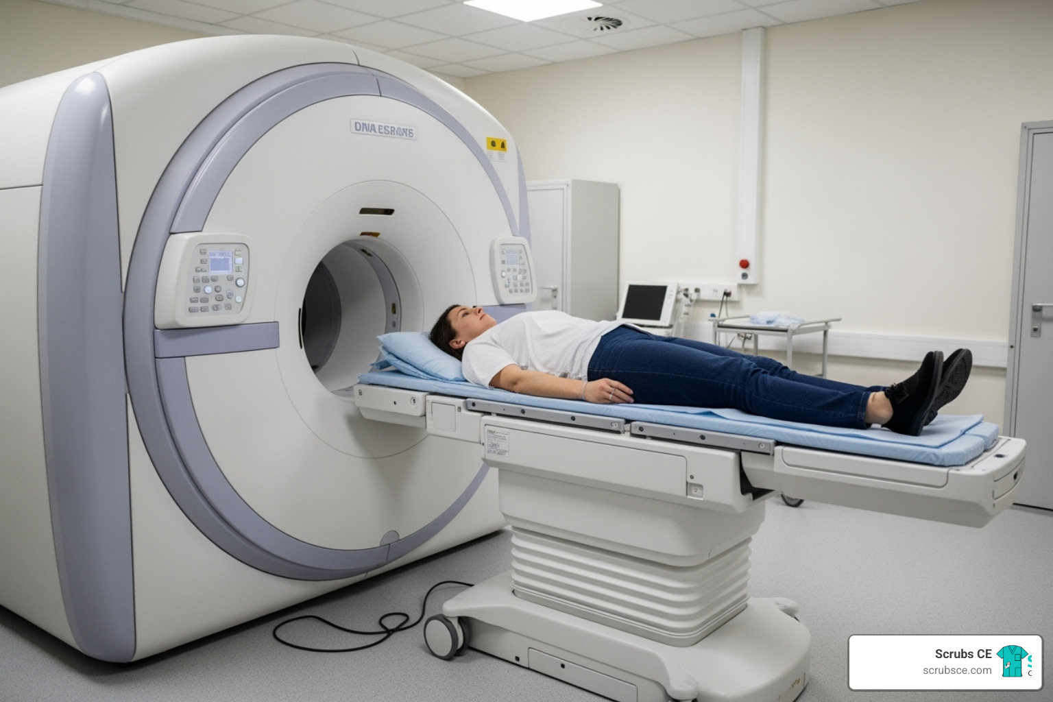
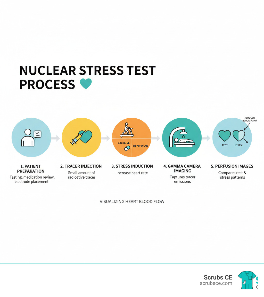
A nuclear medicine myocardial perfusion scan is typically a two-part test comparing blood flow to your heart at rest and during stress. A small amount of a radioactive tracer is injected via an IV, and a special camera (SPECT or PET) tracks where the blood flows. Healthy areas light up, while areas with poor flow appear dim. The entire process takes a few hours but provides a comprehensive picture of your heart’s health.
How to Prepare for Your Scan
Proper preparation is vital for accurate results. Your healthcare team will provide specific instructions, which generally include:
- No caffeine for 24 hours before the scan. This includes coffee, tea, soda, and chocolate, as caffeine can interfere with stress medications.
- Fasting for 4-6 hours before your appointment (water is usually permitted).
- Medication adjustments. You may be asked to temporarily stop certain heart medications, like beta-blockers. Never stop medications without your doctor’s explicit instructions.
- Wear comfortable clothing and athletic shoes for the exercise portion of the test.
During the Scan: Rest vs. Stress
The procedure involves these main steps:
- IV Placement and Rest Scan: An IV line is placed in your arm. The first dose of tracer is injected, and after a waiting period (15-45 minutes), you’ll lie still while a camera takes the resting images.
- Stress Test: Next, your heart is stressed, either through exercise on a treadmill or with a pharmacological stress agent like Lexiscan (regadenoson) that mimics exercise. Your EKG and vital signs are monitored throughout.
- Stress Scan: At peak stress, a second dose of tracer is injected. You will then return to the camera for the second set of images. Comparing the rest and stress images allows doctors to identify areas of ischemia.
For professionals seeking to learn more, more info about Nuclear Medicine CE Courses can provide deeper insights into these protocols.
Potential Risks and Post-Scan Recovery
Nuclear medicine myocardial perfusion scans are very safe. The tracer itself rarely causes side effects. If a stress medication is used, you might feel temporary flushing or a mild headache, but these effects fade quickly.
The radiation exposure is low, comparable to a CT scan. The diagnostic benefit of identifying potentially life-threatening heart disease almost always outweighs this minimal risk.
Recovery is simple. You can resume normal activities immediately. Drinking plenty of fluids will help flush the tracer from your body over the next 24-48 hours. The test has an excellent safety profile, with millions performed worldwide each year.
Interpreting Scan Results and The Role of Nuclear Medicine Myocardial Perfusion
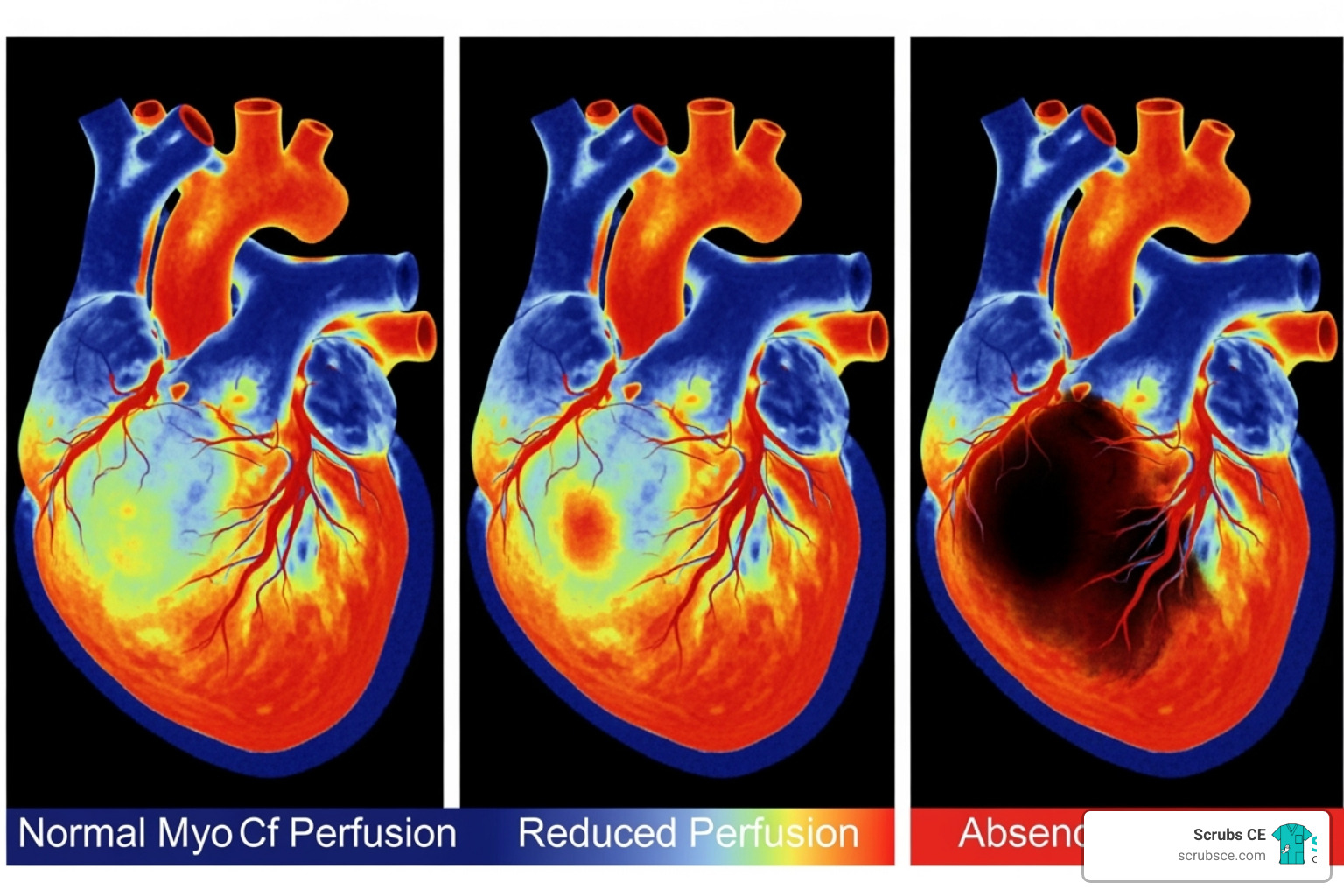
After the scan, a physician analyzes the images by comparing blood flow patterns at rest and during stress. The radioactive tracer highlights areas with good blood flow (“hot spots”), while areas with poor flow appear as “cold spots” or defects.
The key is the comparison between rest and stress images:
- Reversible defects indicate ischemia. A “cold spot” that appears during stress but looks normal at rest signifies a temporary blood flow shortage, typically due to a coronary artery blockage. This condition is often treatable.
- Fixed defects suggest infarction (scar tissue). A “cold spot” that is present on both rest and stress images indicates permanent heart muscle damage from a prior heart attack.
Cardiologists often use a Summed Stress Score (SSS) to quantify the extent and severity of perfusion defects. A higher SSS score correlates with a worse prognosis, making nuclear medicine myocardial perfusion a powerful tool for risk stratification.
What Abnormal Results Mean and Next Steps
An abnormal scan indicates that parts of your heart are not receiving enough blood, which is associated with a significantly higher risk of death or heart attack (11.8% vs. 3.3% for normal scans). However, these results provide a clear roadmap for treatment.
Based on the severity of the findings, next steps may include:
- Medical Therapy: For mild ischemia, lifestyle changes and medications are often the first line of treatment.
- Revascularization: For more significant ischemia, procedures may be recommended to restore blood flow. These include angioplasty and stenting to open narrowed arteries or coronary artery bypass surgery (CABG) for more widespread disease.
Your doctor may also order a coronary angiogram for a more detailed look at the arteries. For more information on imaging standards, professionals can consult Guidance on SPECT Myocardial Perfusion Imaging from the IAEA.
Using MPI to Assess Treatment and Predict Outcomes
The value of nuclear medicine myocardial perfusion extends beyond initial diagnosis. It is used to evaluate the success of interventions like bypass surgery or stenting by confirming that blood flow has been restored. For patients with known CAD, periodic scans can monitor disease progression.
Most importantly, MPI has powerful prognostic value. A normal MPI scan is associated with an excellent prognosis, with a major adverse cardiovascular event rate of just 0.6% per year. This ability to predict outcomes allows clinicians to personalize care, applying aggressive treatment to high-risk patients while providing reassurance to those at low risk.
MPI vs. Other Cardiac Imaging Modalities
While nuclear medicine myocardial perfusion imaging is a cornerstone of cardiac diagnosis, it is part of a larger toolkit. Choosing the right test, or using a multimodality approach, depends on the specific clinical question.
Here’s a comparison of the major cardiac imaging modalities:
- SPECT/PET MPI: Excellent for assessing ischemia and viability. SPECT is widely available, while PET offers higher accuracy (92.6% sensitivity), quantitative blood flow data, and lower radiation. Their main role is functional assessment of blood flow.
- Cardiac MRI (CMR): Offers high accuracy (88% sensitivity, 90% specificity for perfusion) with the major advantage of zero radiation exposure. It provides comprehensive information on heart structure, function, and tissue characterization (e.g., scarring) in a single exam. It is an excellent choice for younger patients or when detailed anatomical and functional data is needed.
- CT Perfusion (CTP): When combined with coronary CT angiography (CCTA), this technique provides both anatomical detail of the arteries and functional perfusion data in one session (86% sensitivity, 92% specificity). This integrated approach is valuable for assessing the significance of a visible blockage, but it involves a moderate to high radiation dose and iodinated contrast.
- Myocardial Contrast Echocardiography (MCE): This ultrasound-based technique is portable, radiation-free, and cost-effective. It offers real-time imaging of perfusion and wall motion at the bedside, with good accuracy (83% sensitivity) and strong agreement with MPI (kappa 0.81). Its quality can be limited by patient body type and operator skill.
Limitations of Nuclear Medicine Myocardial Perfusion
Despite its strengths, MPI is not always the best choice. Key limitations and contraindications include:
- Pregnancy and Breastfeeding: Nuclear imaging is generally avoided. Stress echo or CMR are safer alternatives.
- Severe Respiratory Disease: Patients with severe, uncontrolled asthma or COPD may not be candidates for certain pharmacological stress agents.
- Acute Cardiac Emergencies: Stress testing is unsafe for patients with an ongoing heart attack or unstable angina.
- Obesity: Severe obesity can cause artifacts (especially with SPECT), potentially leading to false-positive results. PET or CMR may be preferred.
- Inadequate Stress: An inadequate exercise test may fail to reveal ischemia, potentially leading to a false-negative result.
- Balanced Ischemia: In rare cases of severe, multi-vessel disease, blood flow may be uniformly reduced, causing the scan to appear deceptively normal.
- Left Bundle Branch Block (LBBB): This conduction abnormality can cause false-positive defects on exercise stress tests. Pharmacological stress is preferred.
Conclusion
Nuclear medicine myocardial perfusion imaging is a powerful diagnostic tool that provides clear, actionable answers about heart health. By visualizing blood flow at rest and under stress, MPI allows clinicians to diagnose coronary artery disease, distinguish treatable ischemia from permanent scar tissue, and assess a patient’s risk for future cardiac events. A normal scan offers powerful reassurance (a 0.6% annual event rate), while an abnormal scan provides a roadmap for life-saving interventions.
For healthcare professionals, mastering these imaging techniques is essential for providing the best patient care. As the field evolves with new tracers and improved cameras, staying current directly translates to better patient outcomes. Understanding the relative strengths of MPI, cardiac MRI, and CT perfusion enables more informed diagnostic strategies.
At ScrubsCE.com, we understand your commitment to excellence. Our convenient, self-paced online courses are designed to fit your busy schedule while delivering the in-depth knowledge you need. Dive deep into complex topics like nuclear medicine myocardial perfusion and earn your continuing education credits on your own time.
Ready to improve your expertise? Explore our comprehensive Nuclear Medicine CE courses and see how staying current can transform your practice.


Recent Comments