
Future-Proofing Your Imaging Department: A Guide to CT Hardware Upgrades
Why CT Tech Hardware Upgrades Matter for Your Imaging Department
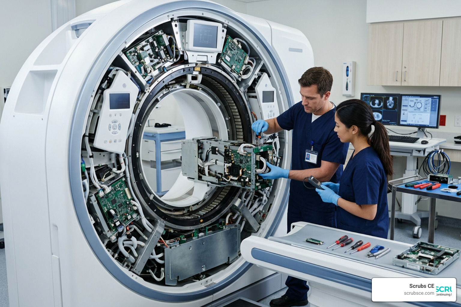
CT tech hardware upgrades are one of the most high-stakes decisions a Radiology department can make — affecting image quality, patient safety, radiation dose, and daily workflow all at once.
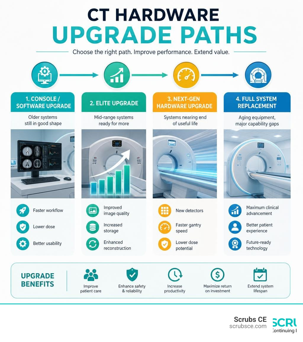
If you’re evaluating your options right now, here’s a quick summary:
| Upgrade Type | Best For | Key Benefit |
|---|---|---|
| Console/software upgrade | Older systems still in good shape | Faster workflow, lower dose |
| Elite upgrade | Mid-range systems (e.g., Optima CT660) | Improved reconstruction, storage |
| Next-gen hardware upgrade | Systems nearing end of useful life | New detectors, gantry speed |
| Full system replacement | Aging equipment, major capability gaps | Maximum clinical advancement |
Radiology departments are under real pressure right now. According to industry data, 73% of departments cite operational efficiency as their top challenge, 35% are dealing with staff shortages, and 35% of studies are repeated daily — often because of image quality issues that better hardware could prevent.
At the same time, buying a brand-new CT scanner isn’t always realistic. That’s where targeted hardware upgrades come in. They can extend the life of existing systems, close technology gaps, and deliver measurable improvements in dose reduction, image quality, and patient throughput — without the full capital cost of a replacement.
The field has moved fast. Advances like photon-counting detectors, dual-source gantry systems, and deep learning reconstruction have fundamentally changed what CT scanners can do. Understanding these changes helps you make smarter investment decisions for your department.
I’m Zita Ewert, and through my work leading continuing education at SCRUBS Continuing Education®, I’ve helped thousands of CT technologists stay current on CT tech hardware upgrades and the clinical knowledge needed to use new systems effectively. In this guide, I’ll walk you through everything you need to evaluate your upgrade options with confidence.
Key Hardware Advancements in Modern CT Systems
When we look at the anatomy of a modern CT scanner, several key hardware components dictate its overall performance: the detector array, the X-ray tube, and the gantry drive system. Upgrading these components directly translates to improvements in spatial resolution (the ability to see tiny structures), temporal resolution (the speed of image capture), and overall patient safety.
Over the last few decades, detector collimation has shrunk from a bulky 13 mm down to an ultra-fine 0.5 mm or less. Simultaneously, X-ray tubes have evolved to deliver up to 120 kW of power with heat dissipation rates exceeding 7 MHU/min. These heavy-duty tubes allow for continuous, high-power scanning without the system needing to pause and cool down—a massive win for trauma centers and high-volume clinics.
Photon-Counting Detectors vs. Energy-Integrating Detectors
The transition from traditional Energy-Integrating Detectors (EIDs) to Photon-Counting Detectors (PCDs) represents a quantum leap in CT technology.
Traditional EIDs rely on a two-step process. First, a scintillator material converts incoming X-ray photons into visible light. Second, a photodiode converts that light into an electrical signal. This indirect conversion has a major drawback: the light scatters within the scintillator layer, which blurs the signal and limits spatial resolution. Additionally, EIDs integrate the energy of all arriving photons over a specific timeframe, meaning they cannot easily distinguish the individual energy levels of the incoming X-rays.
Photon-counting detectors, by contrast, utilize direct conversion. They replace the scintillator material with a semiconductor crystal (such as cadmium telluride). When an X-ray photon hits this crystal, it directly creates pairs of electrical charges that are pulled to electrodes, generating an electrical pulse.
This direct conversion offers massive clinical advantages:
- Zero Light Scattering: Eliminating the scintillator layer drastically improves spatial resolution, allowing us to see microstructures like inner ear bones or fine coronary stents with unprecedented clarity.
- Energy Discrimination: PCDs measure the exact energy of each individual photon. This enables native spectral (dual-energy) imaging on every single scan, without requiring specialized dual-source hardware.
- Electronic Noise Elimination: By setting an energy threshold, PCDs can ignore low-energy electronic noise, resulting in incredibly clean images at significantly lower radiation doses.
Understanding these structural differences is essential for modern technologists. To dive deeper into how detectors and clinical anatomy intersect, check out our course on Computed Tomography for Radiologic Technologists.
Dual-Source Scanners and Gantry Rotation in Cardiac Imaging
In cardiac CT, motion is the ultimate enemy. To capture clear images of a beating heart without motion artifacts, we need exceptional temporal resolution.
Single-source CT systems rely on a single X-ray tube and detector array. To reconstruct an image, the gantry must rotate at least 180 degrees (a half-rotation plus the detector fan angle). Even with lightning-fast gantry rotation times of 270 to 280 milliseconds, the temporal resolution of a single-source scanner is capped at roughly half the rotation time (around 135 to 140 ms). If a patient has a high or irregular heart rate, this is often too slow to prevent blur.
Dual-source CT scanners solve this problem by mounting two X-ray tubes and two corresponding detector arrays at a 90-degree angle on the same gantry. Because the system collects data from two sources simultaneously, it only needs to rotate 90 degrees to acquire a complete 180-degree dataset. This slashes the temporal resolution to one-fourth of the gantry rotation time.
For instance, a dual-source scanner with a 250 ms gantry rotation speed achieves a staggering 66 ms temporal resolution purely through hardware design. This allows for motion-free coronary CT angiography (CTA) and precise myocardial perfusion assessments in a single beat, regardless of the patient’s heart rate. If you are preparing to work with these advanced cardiac configurations, our Computed Tomography 2 course provides an excellent technical foundation.
Strategic Planning for CT Tech Hardware Upgrades

Upgrading an imaging department requires balancing clinical needs with strict capital budgets. Rather than replacing a multi-million dollar CT scanner every time technology advances, we can leverage strategic ct tech hardware upgrades to breathe new life into older systems.
Upgrade Paths for Legacy Systems: Console, Elite, and Next-Gen Options
Major manufacturers have established structured upgrade programs to help facilities extend the lifespan of their equipment. For common platforms like the Revolution CT, Optima CT660, Revolution Maxima, or Revolution EVO, several upgrade tiers are typically available:
- Console and Reconstruction Server Upgrades: This path replaces the operator console and the reconstruction engine with modern, high-speed computing platforms. It increases reconstruction speed, expands local raw data storage, and allows the system to run modern software applications (such as deep learning reconstruction).
- Elite Upgrades: A step up from console upgrades, Elite packages often introduce enhanced hardware components, such as upgraded detector electronics or advanced tube-cooling mechanics, alongside workflow automation tools.
- Next-Gen Hardware Upgrades: This is the most comprehensive upgrade path, where core imaging hardware—such as the X-ray tube, detector modules, or even the gantry drive system—is replaced with next-generation components. This effectively transforms an older system (like an Optima CT660) into a modern clinical workhorse.
By choosing these modular paths, facilities can gain advanced visualization capabilities and faster workflows without the disruptive construction costs of a full room remodel. For those interested in how modular upgrades are executed on-site, you can read more about custom hardware integrations via CTNY Tech On-Site Services: Upgrades.
Choosing Between Console Upgrades and Full System Replacements
Deciding whether to patch up an existing scanner or buy a new one is a classic cost-benefit dilemma.
When evaluating your system, consider these three factors:
- Compatibility and Physical Limits: Standard IT hardware upgrades often hit strict compatibility walls. Just as upgrading consumer PCs can be limited by motherboard chipsets—as detailed in guides like the Upgrade HP 27-CT & 27-CR All-in-One: Complete Guide—medical imaging systems face rigid physical constraints. If your current gantry cannot physically spin faster than 0.5 seconds, or if the detector array cannot handle more than 64 slices, a console upgrade will not change those physical limitations.
- Component Lifecycles: Just as semiconductor transitions impact industrial hardware—such as the Ethernet controller migrations described in this Product Change Notification—medical manufacturers periodically end support for older parts. If your scanner’s core components are designated as “end-of-life,” a full replacement may be the only way to guarantee long-term uptime.
- Implementation Timelines: A console upgrade can often be completed over a weekend, resulting in minimal patient disruption. A full system replacement, however, requires weeks of construction, decommissioning, rigging, and dynamic calibration.
For vintage or highly specialized hardware platforms, keeping legacy systems alive sometimes requires incredibly creative engineering, as seen in historical computing upgrades like the CT60 – Atari Wiki and its associated CTPCI Overview. Fortunately, for medical-grade CT scanners, manufacturers provide highly reliable, certified upgrade paths that guarantee clinical compliance.
Software and Reconstruction Algorithms Complementing Hardware
While hardware determines the raw data a CT scanner can collect, it is the reconstruction software that turns that raw data into diagnostic images. Upgrading your scanner’s computing platform allows you to run modern, mathematically intensive reconstruction algorithms that drastically reduce radiation dose while maintaining—or even improving—image quality.
Historically, CT systems relied on Filtered Back Projection (FBP) to reconstruct images. While FBP is incredibly fast, it assumes that X-ray beams are perfectly parallel and infinitely thin, and that detectors are simple points. Because these assumptions do not match physical reality, FBP produces noisy images when radiation doses are lowered.
Iterative Reconstruction (IR) and Model-Based Iterative Reconstruction (MBIR) take a different approach. They model the complex physics of the X-ray tube, the geometry of the scanner, and the behavior of the detector. The software creates a simulated image, compares it to the raw data, and refines the image over multiple mathematical loops (iterations) until the noise is filtered out.
| Feature | Filtered Back Projection (FBP) | Iterative Reconstruction (IR / MBIR) |
|---|---|---|
| Reconstruction Speed | Extremely fast (near real-time) | Moderate to slow (requires high-power GPUs) |
| Noise Handling | Poor at low radiation doses | Excellent; filters noise mathematically |
| Spatial Resolution | Standard | Significantly enhanced |
| Dose Reduction | Baseline | Up to 60% – 80% lower dose required |
Dose Optimization and Patient Safety in CT Tech Hardware Upgrades
The clinical impact of these software advancements is profound. For example, GE Healthcare’s Veo (a fully model-based iterative reconstruction technology) can achieve clear chest CT images with less than 1 mSv of dose, compared to traditional chest scans that expose patients to 5 to 10 mSv. Similarly, Siemens’ Somatom Perspective utilizes SAFIRE (Sinogram Affirmed Iterative Reconstruction) software to reduce dose by up to 60 percent compared to older FBP techniques.
By combining new detector hardware with modern reconstruction software, imaging departments can safely implement low-dose scanning protocols (such as 70 kVp settings for pediatric or small-stature patients) without sacrificing diagnostic confidence. Keeping up with these safety standards is a core requirement for licensed professionals. To learn more about managing these dose-saving technologies, explore our comprehensive Computed Tomography CE Complete Guide.
Operational Impact: Efficiency, Workload, and Patient Volume
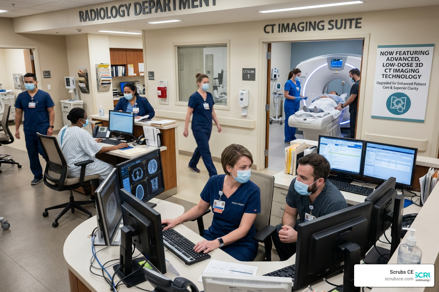
With 73% of Radiology departments identifying operational efficiency as their biggest hurdle, and 35% facing acute staff shortages, ct tech hardware upgrades are no longer just about clinical optics—they are survival tools for busy clinics.
When a scanner is slow, bottlenecks occur. Patients back up in the waiting room, technologists face burnout, and the cost per scan rises. Upgrading your CT hardware directly addresses these operational pain points:
- Fewer Repeat Studies: With 35% of studies repeated daily due to patient motion or poor image quality, faster gantry rotation and better detectors dramatically lower the rate of ruined scans.
- Accelerated Reconstruction Times: Upgraded reconstruction servers process raw data in seconds, allowing technologists to verify image quality before the patient even leaves the table.
- Simplified Protocols: Modern user interfaces automate protocol selection based on the patient’s body habitus, reducing setup times and minimizing human error.
Streamlining Workflow with Smart CT Tech Hardware Upgrades
Operational efficiency also extends to auxiliary hardware, such as contrast injectors. Modern “smart” CT injector systems integrate directly with the scanner’s console. These systems capture patient-enriched data, automatically adjust contrast volume based on patient weight, and coordinate saline chase administration.
Using a saline chase immediately after the contrast bolus pushes the contrast medium forward, ensuring optimal vascular opacification with less total contrast volume. This not only improves image quality but also reduces contrast costs and lessens the excretory burden on the patient’s kidneys.
By automating these steps, technologists can focus on patient care and safety rather than manual calculations. To stay competitive in these clinical environments, check out our resource on the Best CE for CT Technologists to find courses that focus on modern clinical workflows.
Advanced Clinical Applications Enabled by Hardware Upgrades
Investing in ct tech hardware upgrades unlocks clinical pathways that were once completely impossible or required invasive procedures.
- Spectral and Dual-Energy Imaging: By utilizing detectors capable of distinguishing different X-ray energy levels, clinicians can perform material decomposition. This allows them to virtually subtract iodine from an image (creating a virtual unenhanced scan without re-scanning the patient) or isolate uric acid crystals to definitively diagnose gout.
- Quantitative Plaque Analysis: High-resolution detectors allow software to characterize coronary plaques beyond simple stenosis percentages. Technologists can identify high-risk plaque features—such as low-attenuation cores, positive remodeling, and the “napkin-ring sign”—which are strong predictors of acute coronary syndrome (ACS).
- Noninvasive Fractional Flow Reserve (FFRCT): Historically, determining whether a coronary artery blockage was hemodynamically significant required an invasive cardiac catheterization to measure Fractional Flow Reserve (FFR). Today, combining high-resolution CT data with advanced computational fluid dynamics allows for noninvasive FFRCT. Clinical trials (like the NXT trial) have demonstrated that FFRCT achieves a per-patient sensitivity of 86% and an accuracy of 81% in identifying lesions causing ischemia, safely keeping patients out of the cath lab.
Frequently Asked Questions about CT Upgrades
What are the primary benefits of photon-counting detectors?
Photon-counting detectors offer unprecedented spatial resolution by converting X-ray photons directly into electrical signals, eliminating the light-scattering blur of traditional scintillators. They also allow for native spectral imaging on every scan and significantly reduce electronic noise, enabling high-quality images at much lower radiation doses.
How do hardware upgrades improve cardiac CT imaging?
Upgrades like dual-source configurations and faster gantry rotation times drastically improve temporal resolution (down to 66 ms). This allows the scanner to capture motion-free images of the coronary arteries even in patients with high or irregular heart rates, reducing the need for beta-blockers before a scan.
When should a facility choose a console upgrade over a full replacement?
A console upgrade is ideal when the scanner’s physical hardware (gantry, tube, and detectors) is still in excellent condition, but the computing system is too slow to support modern iterative reconstruction or advanced workflow software. It is a highly cost-effective way to extend the system’s life by 3 to 5 years.
Conclusion
Future-proofing your imaging department doesn’t always require purchasing a brand-new CT scanner. Through strategic ct tech hardware upgrades, facilities can achieve state-of-the-art spatial resolution, dramatic dose reductions, and streamlined workflows at a fraction of the cost of a full replacement.
As these technologies continue to evolve, staying updated on how to operate them is essential for professional development. Under the guidelines of the AMERICAN REGISTRY OF RADIOLOGIC TECHNOLOGISTS® (ARRT®) and the ASRT®, technologists must maintain continuous education to ensure patient safety and clinical excellence.
At Scrubs CE (ScrubsCE.com), we provide convenient, self-paced, and affordable online courses designed to help you meet your licensure requirements and master these cutting-edge imaging technologies. Ready to take the next step in your professional journey? Explore our Computed Tomography Certification courses today and keep your career moving forward.