
Fast-Track Your CMR Competency with Hands-On Clinical Training
Core Frameworks for Cardiac MRI Training: Levels I, II, and III Standards
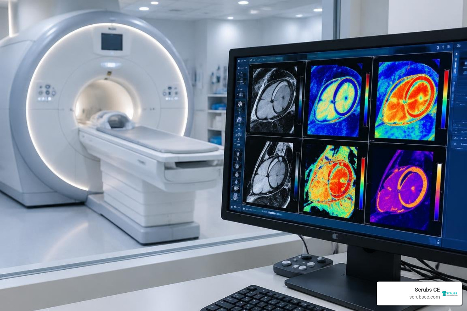
Cardiovascular magnetic resonance (CMR) has evolved from a specialized research tool into an essential clinical modality. To ensure consistent quality and patient safety, professional bodies like the Society for Cardiovascular Magnetic Resonance (SCMR) and the American College of Cardiology (ACC) have established clear diagnostic benchmarks. These frameworks define three progressive tiers of competency.
Surveys highlight an ongoing educational gap: approximately 64% of responding practitioners report having no formal CMR training, and 55% lack official verification of competence. Furthermore, while 68% felt their primary residency or fellowship met basic educational needs, 84% expressed a strong need for expanded training opportunities. Structured competency levels provide the clear path forward.
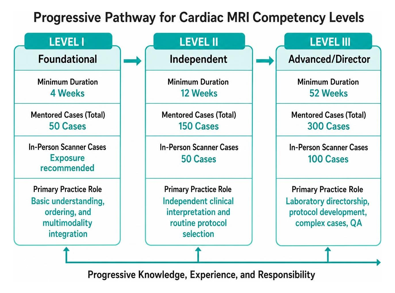
| Competency Level | Minimum Duration | Mentored Cases (Total) | In-Person Scanner Cases | Primary Practice Role |
|---|---|---|---|---|
| Level I (Foundational) | 4 Weeks | 50 Cases | Exposure recommended | Basic understanding, ordering, and multimodality integration |
| Level II (Independent) | 12 Weeks | 150 Cases | 50 Cases | Independent clinical interpretation and routine protocol selection |
| Level III (Advanced/Director) | 52 Weeks | 300 Cases | 100 Cases | Laboratory directorship, protocol development, complex cases, QA |
SCMR Guidelines and Case Volume Requirements
The SCMR training guidelines emphasize that achieving true competency requires both cumulative case volumes and active, supervised participation. Level I provides the foundation, introducing basic physics, anatomy, indications, and imaging contraindications over a 4-week dedicated period.
Level II requires 12 weeks of dedicated training and 150 mentored cases (with active scanner presence for at least 50). This tier qualifies a specialist to interpret routine clinical studies independently. For those aiming to lead an imaging facility, Level III requires a full year of training and 300 mentored cases (with 100 on-site at the scanner). The SCMR position statement on a competency-based CMR curriculum notes that logbooks must document hands-on protocol setup, real-time troubleshooting, and formal post-processing to ensure trainees develop true diagnostic skill.
Navigating Pathways for Cardiology and Radiology Specialists
Cardiologists and radiologists bring distinct, complementary strengths to CMR. Cardiology trainees often possess deep insights into hemodynamics, electrophysiology, and bedside clinical management, but they may need more structured instruction in advanced pulse sequences and artifact physics. Radiologists usually have strong foundations in MR physics, cross-sectional anatomy, and non-cardiac incidental findings, but they may require deeper exposure to cardiovascular pathophysiology.
Harmonizing these curricula requires interdisciplinary collaboration. Programs that run joint imaging services allow fellows from both specialties to learn together. Understanding technical scanner operations and collaborating with certified technologists—whose core clinical duties are outlined in this guide on MRI technologist job description and key responsibilities—ensures smooth clinical workflows and accurate image acquisition.
Essential Technical Competencies: MRI Physics, Safety, and Clinical Interpretation
Achieving independent proficiency requires mastering both technical and clinical aspects of CMR. Unlike static imaging, cardiac MRI demands dynamic synchronization with cardiac and respiratory motion.
Trainees must understand how radiofrequency pulses, gradient switching, and k-space trajectory design affect diagnostic quality. Balancing signal-to-noise ratio (SNR), temporal resolution, and spatial resolution is essential when evaluating moving cardiac structures. Reviewing foundational concepts through resources on MRI basics and foundational physics helps clinicians troubleshoot artifacts such as phase wrap, metallic distortion, and arrhythmia-induced ghosting.
Advanced Cardiac MRI Training in Protocol Optimization and Tissue Characterization
Standard cine imaging using steady-state free precession (SSFP) serves as the reference standard for ventricular volume, ejection fraction, and wall-motion assessment. Beyond functional evaluation, tissue characterization is where CMR excels:
- T1 and T2 Parametric Mapping: Quantifies native myocardial relaxation times to detect diffuse interstitial fibrosis, amyloid deposition, and acute myocardial edema without relying solely on relative visual contrasts.
- Late Gadolinium Enhancement (LGE): The established benchmark for assessing myocardial viability, distinguishing ischemic subendocardial infarction from non-ischemic patterns like mid-wall or subepicardial fibrosis in myocarditis and genetic cardiomyopathies.
- Extracellular Volume (ECV) Quantification: Combines pre- and post-contrast hematocrit-adjusted T1 maps to assess expansion of the extracellular matrix.
Trainees can explore these advanced pulse sequences through specialized symposia like the ISMRM cardiovascular MRI educational sessions.
Contrast Safety and Hemodynamic Assessment in Clinical Practice
Administering Gadolinium-Based Contrast Agents (GBCAs) requires careful attention to patient safety. Modern macrocyclic formulations provide exceptional kinetic stability and low dissociation rates, minimizing the risk of Nephrogenic Systemic Fibrosis (NSF) in patients with renal impairment. High-relaxivity agents, such as gadopiclenol, achieve strong contrast enhancement at lower total gadolinium doses, which is especially useful during dual-bolus stress perfusion exams.
Hemodynamic evaluation also includes phase-contrast velocity encoding (VENC) and 4D flow imaging to assess valvular regurgitation, intracardiac shunts, and complex aortic hemodynamics. Technologists and imaging professionals can expand their understanding of these clinical protocols through CE for MRI technologists.
Modern Educational Modalities: Virtual Simulators, Compact Courses, and AI Integration
Traditional training pathways often faced bottlenecks due to limited scanner time and regional variations in case complexity. Modern educational strategies help bridge these gaps by combining interactive digital tools with hands-on clinical practice.
Digital Strategies and Simulator Platforms for Cardiac MRI Training
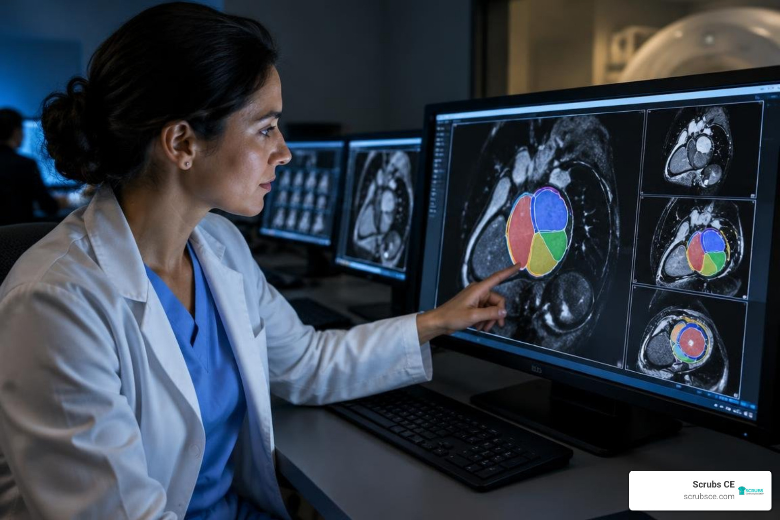
Interactive simulation software allows trainees to operate virtual scanner consoles, adjust imaging planes (such as 2-chamber, 4-chamber, and short-axis views), and modify parameters to manage arrhythmias without using valuable clinical magnet time. Platforms like the Siemens Healthineers virtual cardiac training offer structured environments to practice challenging scenarios before working with live patients. These tools, paired with structured online MRI courses, help learners review diverse, curated pathological cases on demand.
Fellowship Models, Global Compact Courses, and AI Workflows
For clinicians who cannot take a full one-year leave for advanced fellowship training, modular options provide a flexible alternative:
- Intensive 5-Day Compact Courses: Offer immersive lectures, protocol demonstrations, and 50 to 100 guided case reviews to meet Level I or partial Level II requirements.
- High-Volume Clinical Fellowships: Short-term mini-fellowships (1 to 3 months) at tertiary institutions provide exposure to high case volumes, such as the thousands of annual scans performed at centers like Pisa or the Royal Brompton Hospital.
- AI-Driven Workflows: Deep learning algorithms now automate contouring, volume calculation, flow analysis, and image reconstruction. Trainees who understand how to review and validate these automated tools can interpret scans more efficiently as demand grows, a trend explored in our review of the MRI tech job outlook and career growth in 2026.
Certification, Institutional Credentialing, and Career Advancement
Completing an educational curriculum is the first step; obtaining formal credentials validates clinical competence for institutional privileging and independent billing.

Formal credentialing protects patient safety and confirms that an imaging specialist meets national practice standards. For technologists, maintaining primary credentials from the AMERICAN REGISTRY OF RADIOLOGIC TECHNOLOGISTS® is essential. Our ARRT® MRI registry review preparation provides a focused resource for mastering registry examinations administered by the ARRT®.
CBCMR Certification and Intersocietal Accreditation Commission Standards
The Certification Board of Cardiovascular Magnetic Resonance (CBCMR) offers a rigorous examination for physicians. Candidates can qualify through formal Level II/III fellowship tracks or an established practice pathway (which requires maintaining an active CMR practice for 3 of the previous 5 years with at least 300 interpreted studies).
For institutional facility accreditation through the Intersocietal Accreditation Commission (IAC), interpreting physicians must document formal Level II training with 150 supervised cases, or demonstrate informal training within an accredited facility alongside 150 cases and 150 hours of relevant Category 1 Continuing Medical Education (CME).
Maintaining Continuing Competency and Practice Verification
To maintain diagnostic accuracy, imaging laboratories and practitioners should follow structured quality-assurance protocols:
- Complete at least 30 to 60 hours of cardiac-specific Category 1 CME or CE every two to three years.
- Participate in regular interdisciplinary clinical review conferences alongside Radiology and cardiology colleagues.
- Conduct quarterly internal peer-review audits, cross-checking CMR diagnoses against surgical findings, catheterization results, and histopathology.
- Maintain a balanced case mix covering stress viability, non-ischemic myopathies, flow quantification, and adult congenital heart disease.
Frequently Asked Questions About Cardiac MRI Training
What are the case volume requirements for Level II cardiac MRI competency?
Under SCMR and ACC training guidelines, Level II competency requires interpreting at least 150 mentored cases under the supervision of a Level III expert. Trainees must also be physically present at the scanner console for a minimum of 50 acquisitions, alongside 12 dedicated weeks of training, to qualify for independent clinical reporting.
How long does it take to complete independent cardiac MRI training?
Achieving independent Level II competency typically takes 3 to 6 months of dedicated fellowship training or structured modular rotations. Advanced Level III competency, which prepares practitioners for laboratory directorship and academic leadership, requires a full 12-month dedicated imaging fellowship.
Can cardiac MRI training and certification be completed through virtual or hybrid courses?
Virtual simulators and digital courses can fulfill didactic lecture hours, CME requirements, and remote case-interpretation quotas. However, official Level II and Level III certifications still require on-site scanner console time to master patient positioning, ECG gating, contrast timing, and artifact troubleshooting.
Conclusion
Building real competency in cardiac MRI requires balanced dedication to foundational physics, protocol optimization, and guided clinical case review. As clinical guidelines increasingly recommend CMR as a primary diagnostic tool for complex cardiac disease, skilled practitioners and imaging professionals remain in high demand.
At Scrubs CE, we support your ongoing professional development by providing high-quality, flexible, and affordable e-learning opportunities. Whether you are building foundational knowledge or completing mandatory licensure requirements, explore our comprehensive MRI continuing education credits to advance your clinical expertise with self-paced courses and instant certificates.