

Why Anatomical Imaging Examples Matter for Every Imaging Professional
Anatomical imaging examples give clinicians, students, and imaging professionals a clear window into the human body — without a single incision. From a simple chest X-ray to a 3D synchrotron scan of an intact human brain, these techniques have transformed how we diagnose disease, plan surgery, and understand the body at every scale.
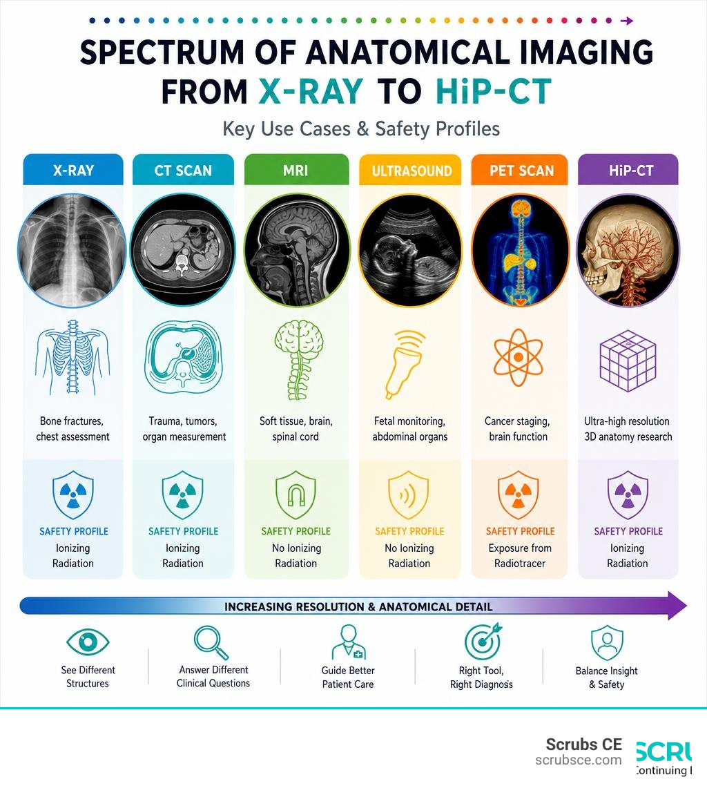
Here is a quick overview of the most common anatomical imaging modalities and what each one is best used for:
| Modality | How It Works | Best Used For |
|---|---|---|
| X-Ray | Ionizing radiation passes through tissue | Bone fractures, chest assessment |
| CT Scan | X-rays captured at multiple angles | Trauma, tumors, organ measurement |
| MRI | Magnetic fields + radio waves | Soft tissue, brain, spinal cord |
| Ultrasound | High-frequency sound waves | Fetal monitoring, abdominal organs |
| PET Scan | Radiotracer detects metabolic activity | Cancer staging, brain function |
Each modality shows a different “version” of the body. No single technique does everything. Knowing which tool fits which clinical question is a core skill for any imaging professional — and it starts with seeing real examples.
I’m Zita Ewert, and as the force behind SCRUBS Continuing Education®, I’ve spent years helping radiologic technologists and imaging professionals stay current on topics like anatomical imaging examples through practical, accredited coursework approved by the AMERICAN REGISTRY OF RADIOLOGIC TECHNOLOGISTS® (ARRT®). In this guide, we’ll walk through every major modality, real-world clinical cases, and the cutting-edge research reshaping what anatomical imaging can do.
Simple anatomical imaging examples word guide:
Anatomical vs. Functional Imaging: Key Differences
When we look at medical imaging, we generally divide the technologies into two main families: anatomical imaging and functional imaging.
- Anatomical imaging focuses on the physical structures, shape, density, and spatial relationships of body tissues. It answers the question: What does it look like, and where is it? Examples include finding a bone fracture on an X-ray or mapping a brain tumor’s boundaries using a high-resolution MRI.
- Functional imaging looks at the physiological and metabolic processes happening inside those structures. It answers the question: How is it working? This includes measuring blood flow, oxygen consumption, or glucose metabolism, such as detecting highly active cancer cells with a PET scan.

In modern medicine, these two fields are no longer isolated. Hybrid systems combine both types of data into a single, comprehensive view. By merging anatomical and functional imaging, systems like PET-CT and PET-MRI allow clinicians to see exactly where a metabolic change is occurring in the physical structure of an organ. This dual perspective is incredibly valuable for oncology, cardiology, and neurology.
| Feature | Anatomical Imaging | Functional Imaging |
|---|---|---|
| Primary Focus | Physical structure, boundaries, and tissue density | Metabolic activity, blood flow, and chemical processes |
| Typical Modalities | X-Ray, CT, MRI, Ultrasound | PET, fMRI, SPECT |
| Clinical Value | Identifying structural damage, tumors, and fractures | Detecting early cellular changes and monitoring organ function |
Core Modalities and Anatomical Imaging Examples
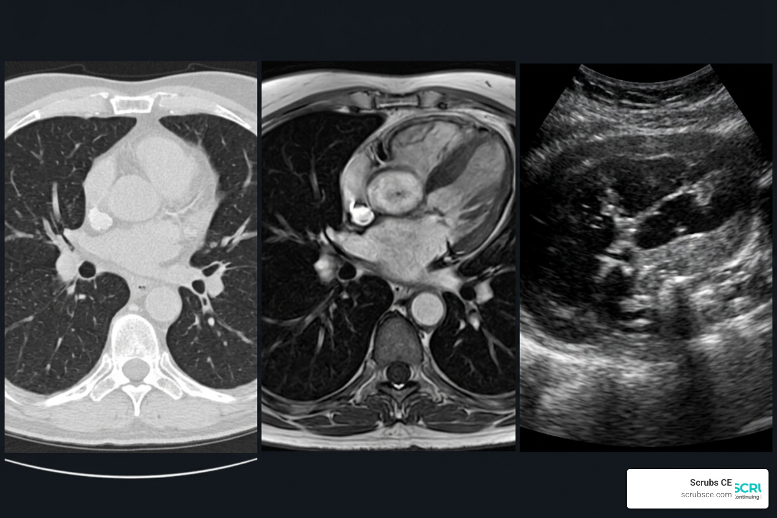
To truly understand how these systems work in clinical practice, we need to look at specific anatomical imaging examples across the core modalities. If you want to dive deeper into how these concepts apply to professional practice, our Anatomy Radiology Book Essentials Top Texts for Diagnostic Imaging guide is an excellent starting point.
X-Ray and Computed Tomography (CT)
X-rays are the oldest and most common form of medical imaging. They use high-energy electromagnetic radiation with a short wavelength to pass through the body. Dense tissues, like bone, block the radiation and appear white, while softer tissues allow the rays to pass through and appear dark.
Computed Tomography (CT) takes this concept to the next dimension. Instead of a single flat image, a CT scanner rotates 360 degrees around the patient, taking multiple X-ray measurements that a computer reconstructs into cross-sectional “slices.” Modern helical CT scans can capture these slices rapidly, which is essential in emergency trauma settings where every second counts.
- Key Clinical Applications: X-rays are perfect for assessing bone fractures, dental anatomy, and chest conditions like pneumonia. CT scans are the gold standard for trauma imaging, identifying internal bleeding, and measuring the precise size of abdominal tumors down to a millimeter.
- Safety & Cost Considerations: While X-rays are inexpensive and fast, CT scans expose patients to a dose of ionizing radiation many times higher than standard X-rays. Because of this, CT scans should not be performed repeatedly unless medically necessary. To learn more about positioning and radiation safety, check out The ABCs of X-ray Physics Anatomy and Perfect Positioning.
Magnetic Resonance Imaging (MRI) and Ultrasound
Magnetic Resonance Imaging (MRI) uses strong magnetic fields and radiofrequency waves to excite hydrogen atoms in the body’s water molecules. As these atoms return to their normal state, they emit radio signals that are captured and turned into highly detailed images.
Ultrasound, on the other hand, relies on high-frequency sound waves. A transducer sends sound waves into the body, which bounce off internal tissues. The returning echoes are converted into real-time moving images.
- Key Clinical Applications: MRI provides unmatched soft-tissue resolution, making it the preferred choice for brain, spinal cord, and joint imaging. For example, clinicians use MRI to trace the intricate paths of cranial nerves, as shown in the Anatomy of the cranial nerves and brainstem: annotated MRI | e-Anatomy module. It is also excellent for complex joints, which you can explore in detail through the Wrist on 3T MR and 3D pictures: normal anatomy | e-Anatomy reference. Ultrasound is highly valued for real-time applications, such as monitoring fetal development during pregnancy or performing bedside assessments for internal bleeding.
- Safety & Cost Considerations: Ultrasonography does not expose patients to radiation, radiopharmaceuticals, or magnetic fields, meaning there are no known medical risks. MRI is also free of ionizing radiation, but the scans can take up to 30 minutes and are incredibly noisy, which can cause patient anxiety. Additionally, patients with metallic implants (like pacemakers) may not be able to undergo an MRI due to the strong magnetic fields.
Advanced 3D and Hierarchical Organ Imaging
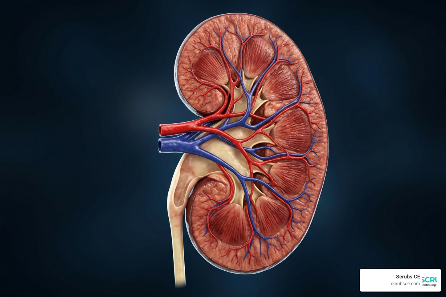
As imaging technology advances, we are moving far beyond flat 2D slices. Today, researchers can capture entire organs in three dimensions, bridging the gap between macroscopic anatomy (what we can see with the naked eye) and microscopic histology (what we see under a microscope).
Hierarchical Phase-Contrast Tomography (HiP-CT) as an Anatomical Imaging Examples Pioneer
A major breakthrough in this area is Hierarchical Phase-Contrast Tomography (HiP-CT). Developed using high-energy synchrotron radiation at the European Synchrotron Radiation Facility (ESRF), this technology utilizes the Extremely Brilliant Source (EBS), which provides a 100-fold increase in X-ray brilliance compared to older synchrotrons.
HiP-CT allows scientists to perform hierarchical imaging of intact human organs. This means they can scan a whole organ at a baseline resolution and then zoom in to cellular-level detail without physically cutting or damaging the tissue. You can read the foundational research on this technology in the paper Imaging intact human organs with local resolution of cellular structures using hierarchical phase-contrast tomography | Nature Methods.
- Unprecedented Resolution: HiP-CT can scan a whole human organ at 25 µm per voxel, zoom in to 6.5 µm, and then achieve an incredible local resolution of 1.3 to 2.5 µm per voxel.
- Scan Times: A whole brain scan at 25 µm per voxel takes about 16 hours, while an intact kidney can be scanned in approximately 3.5 hours.
- Bridging the Gap: Traditional light microscopes magnify up to 1,000x, and electron microscopes (like SEM and TEM) can magnify up to 500,000x or 10,000,000x respectively, but they require cutting the tissue into tiny, thin slices. HiP-CT provides a non-destructive way to view these structures in their native 3D environment.
Real-World Research and Veterinary Anatomical Imaging Examples
These high-resolution imaging techniques are also transforming veterinary medicine, comparative anatomy, and research ethics.
- Reducing Animal Testing: Historically, researchers had to use live animals to study anatomical development over time. Today, tools like the Göttingen Minipigs MR and CT Imaging Atlas allow scientists to study organ growth and structural changes virtually. This directly supports the “3R” principles (Replacement, Reduction, and Refinement) in scientific research.
- Marine Mammal Forensics: In veterinary pathology, post-mortem CT (PMCT) has become an invaluable tool. For example, the open-access database Cet.CT-Bank: A Postmortem Computed Tomography Imaging Data of Stranded Cetaceans from the Canary Islands | Scientific Data provides researchers with 3D anatomical data of stranded dolphins and whales, helping them investigate causes of death without immediate invasive dissection.
Emerging Trends and Quantitative Analysis in Anatomical Imaging Examples
The future of medical imaging lies in turning visual pictures into objective, quantitative data. Instead of simply looking at a scan, modern software can measure, count, and analyze anatomical structures automatically.
Synchrotron Radiation and Phase-Contrast Tomography
Conventional clinical CT scanners rely on X-ray absorption (how much radiation is blocked by tissue). Synchrotron-based phase-contrast imaging, however, measures how the X-ray wave shifts as it passes through different tissues. This provides much higher contrast in soft tissues, allowing for “virtual histology” of whole organs without the need for chemical stains or physical slicing.
AI-Assisted Interpretation and Quantitative Morphometry
Artificial Intelligence (AI) and deep learning algorithms are now used to perform automated segmentations of complex organs. For example, researchers can use these tools to map the dynamic movement of internal organs in real time, as demonstrated in Fig. 5: Label-free visualization of whole-trunk dynamics in 3D-PanoPACT. | Nature Communications.
These technologies allow for incredibly precise quantitative analysis:
- Glomerular Counting: In a kidney scan of a 94-year-old female, HiP-CT was used to estimate the total number of glomeruli (the kidney’s filtering units) at approximately 310,000. This is highly accurate and falls well within the expected physiological range.
- Volume Measurements: The mean glomerular volume was measured at 5.05 ± 0.09 × 10⁻³ mm³, which aligns closely with traditional stereological and MRI analyses.
- Pathology Assessment: AI can analyze 3D lung scans to quantify the exact volume of alveolar obstruction, septal thickening, and capillary damage in patients with severe respiratory diseases, including COVID-19.
If you are currently studying anatomy or preparing for an exam, practicing with visual resources is one of the best ways to build your spatial understanding. We recommend using resources like the Anatomy Coloring Workbook Test Only Emailed to reinforce your knowledge of these complex structures.
Frequently Asked Questions about Anatomical Imaging
What is the safest anatomical imaging modality?
Ultrasonography is widely considered the safest anatomical imaging modality because it does not use ionizing radiation, magnetic fields, or radiopharmaceuticals. There are no known medical risks associated with diagnostic ultrasound, making it the gold standard for monitoring pregnancy. MRI is also very safe because it avoids radiation, but patients must be screened for metallic implants, and the loud, enclosed environment can be challenging for some. If you are looking to learn more about basic anatomy safety and terminology, the Anatomy & Physiology for Dummies 3rd Ed CH Test Only E-mailed course is a great resource.
Why is CT preferred over MRI in emergency trauma cases?
CT is preferred in emergency settings primarily because of its speed and accessibility. A modern helical CT scan can image a patient’s entire body in less than a minute, whereas an MRI scan can take 30 minutes or longer. Additionally, CT is exceptionally good at showing acute bone fractures and fresh bleeding, which are critical to identify immediately in trauma patients.
How does HiP-CT bridge the gap between anatomy and histology?
HiP-CT bridges this gap by providing high-resolution 3D imaging across multiple scales. It allows researchers to view a whole, intact organ (macroscopic anatomy) and then zoom in to resolve individual cells (microscopic histology) down to 1.3 µm, all without physically cutting, staining, or damaging the tissue.
Conclusion
From standard diagnostic X-rays to cutting-edge 3D synchrotron scans, anatomical imaging examples show us how rapidly our ability to visualize the human body is evolving. For radiologic technologists, sonographers, and other imaging professionals, staying up-to-date on these technologies is key to delivering excellent patient care and advancing your career.
At SCRUBS Continuing Education®, we are dedicated to providing high-quality, self-paced online courses with instant certificates to help you meet your professional development and state licensing requirements. Our courses are designed to align with the standards of the AMERICAN REGISTRY OF RADIOLOGIC TECHNOLOGISTS® (ARRT®) and the ASRT®.
Ready to expand your clinical knowledge and earn your CE credits? Explore our complete Anatomy for Radiographers course catalog today!