

What Is Pathological Anatomy?
The pathological anatomy definition is the medical study of disease-related structural changes in cells, tissues, and organs. Also called anatomic pathology or, historically, morbid anatomy, it helps pathologists identify what disease is present, how extensive it is, and sometimes how it may respond to treatment.
In practical terms, pathological anatomy examines samples such as biopsies, surgical specimens, cells collected by needle aspiration or screening tests, and tissues from autopsies. A pathologist combines what can be seen with the naked eye, under a microscope, and through specialized laboratory tests to turn a specimen into a useful diagnostic report.
This field is a core part of modern medicine because many important changes cannot be confirmed by symptoms, imaging, or blood tests alone. In cancer care especially, tissue findings can establish the tumor type, assess surgical margins, estimate prognosis, and identify biomarkers that may guide therapy.
I am Zita Ewert, a continuing-education leader for imaging professionals who focuses on clear, practical learning for busy healthcare teams. Understanding the pathological anatomy definition helps connect diagnostic imaging, tissue diagnosis, and the clinical decisions that follow.
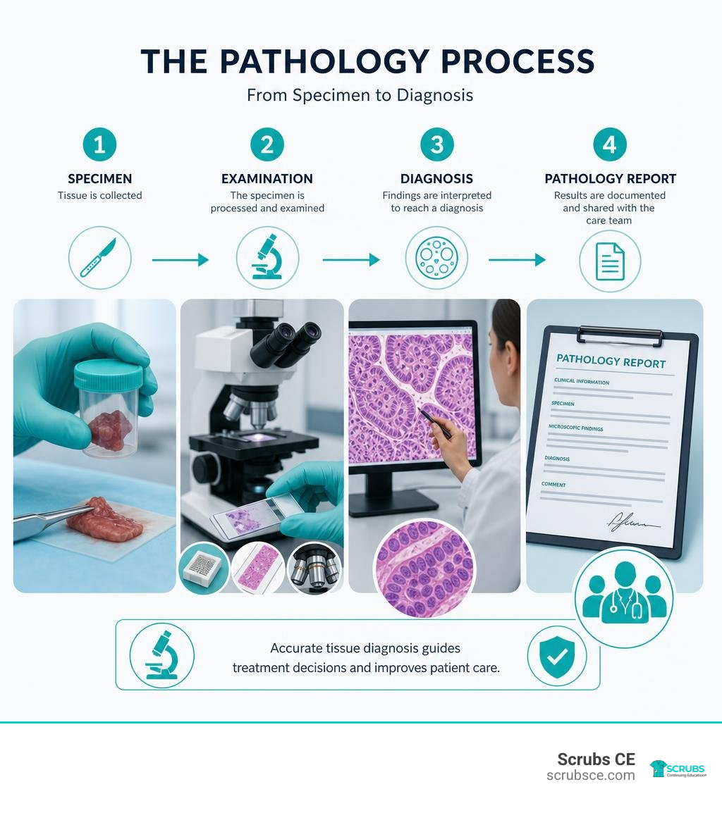
Simple guide to pathological anatomy definition:
Understanding the Pathological Anatomy Definition and Its Scope
To truly grasp the formal pathological anatomy definition, we must examine how it bridges fundamental biological science and frontline clinical care. At its root, pathology stems from the Greek word pathología, which translates to “the study of suffering.” Pathological anatomy focuses specifically on the physical, tangible footprint that suffering leaves behind within the human body.
When a disease takes hold, it alters biological structures across multiple levels—from macroscopic changes visible on an organ’s surface down to microscopic cellular architecture and molecular alterations. In our clinical workflows, we separate the study of disease into two key concepts:
- Etiology: The underlying cause or biological trigger of a disease (such as a viral infection, genetic mutation, or toxic exposure).
- Pathogenesis: The stepwise cellular, chemical, and structural mechanism through which that initial cause develops into full-blown clinical disease.
Pathological anatomy acts as the physical map of pathogenesis. By evaluating tissue architecture and cellular morphology, pathologists trace how normal anatomy transforms into diseased tissue. Whether evaluating a colorectal polyp or analyzing end-stage pulmonary fibrosis—where normal lung tissue degrades into a classic macroscopic “honeycomb” appearance—pathological anatomy provides the baseline ground truth for clinical diagnosis.
For additional reference on standard medical definitions, you can consult this dictionary entry on pathologic anatomy.

Formal Pathological Anatomy Definition vs Clinical Pathology
In modern hospital settings, diagnostic pathology is broadly divided into two major disciplines: anatomical pathology and clinical pathology. While both share the ultimate goal of accurate patient diagnosis, their daily operations, specimen types, and analytical methods differ significantly.
Anatomical pathology centers on the structural and morphological analysis of solid tissues and cellular samples. Pathologists specializing in this branch evaluate tissue biopsies, major surgical resections, fine-needle aspirates, and post-mortem autopsy tissues. They inspect specimens grossly with the naked eye, cut microscopic sections, apply specialized stains, and evaluate cellular patterns under light microscopes or digital screens.
Conversely, clinical pathology focuses primarily on the laboratory analysis of bodily fluids and biochemical markers. Clinical pathologists oversee departments such as clinical chemistry, medical microbiology, hematology, immunology, and blood banking. They analyze blood serum, urine, cerebrospinal fluid, and bone marrow aspirates using automated analyzers to measure cell counts, electrolyte levels, hormone concentrations, and microbial cultures.
| Feature | Pathological (Anatomical) Anatomy | Clinical Pathology |
|---|---|---|
| Primary Specimen Types | Solid tissue biopsies, surgical resections, cytology smears, autopsies | Blood, urine, cerebrospinal fluid, plasma, body secretions |
| Core Analytical Focus | Microscopic cell architecture, tissue morphology, immunophenotyping | Chemical concentrations, cell counts, microbial cultures, fluid dynamics |
| Key Equipment | Microtomes, light/fluorescence microscopes, tissue processors | Automated blood analyzers, centrifuges, mass spectrometers, culture incubators |
| Clinical Objective | Confirm tissue diagnosis, tumor grading, staging, and surgical margin status | Monitor metabolic function, detect systemic infections, measure biomarkers |
Together, these twin pillars ensure that patient care relies on complete laboratory insights—combining structural tissue analysis with fluid chemistry.
Historical Evolution: From Morgagni to Virchow
Pathological anatomy did not emerge fully formed; it evolved over centuries of scientific curiosity, technological breakthroughs, and shifts in medical philosophy. Before the scientific revolution, ancient medicine relied heavily on humoral theory—the belief that disease arose from an imbalance among four bodily humors (blood, phlegm, black bile, and yellow bile).
The shift toward modern anatomical investigation gained significant momentum in 1543, when Andreas Vesalius performed direct human dissections to challenge ancient anatomical misconceptions. However, the modern foundation of pathological anatomy as a systematic discipline was laid in the 18th century by Giovanni Battista Morgagni. In his landmark 1761 publication, De Sedibus et Causis Morborum per Anatomen Indagatis (“On the Seats and Causes of Diseases Investigated by Anatomy”), Morgagni systematically correlated clinical symptoms observed during a patient’s life with structural organ abnormalities discovered during post-mortem autopsies.
During the 19th century, medical centers across Europe—particularly the famous Vienna School—embraced prosection and post-mortem examination. But the ultimate paradigm shift occurred when optics and microtechnology converged.
Rudolf Virchow, widely celebrated as the father of modern cellular pathology, published Cellularpathologie in 1858. Virchow famously posited omnis cellula e cellula (“every cell arises from another cell”) and demonstrated that disease is not simply an imbalance of body fluids or organ-wide dysfunction, but rather stems from structural and functional alterations at the cellular level.
The historical transmission of these medical concepts is thoroughly detailed in this guide on Pathoanatomy Definition.
From early macroscopic cadaver prosection to Virchow’s microscopic breakthroughs, pathological anatomy transformed medicine from speculative philosophy into an exact, structural science.
Key Subspecialties and Specimen Types
As modern medicine has grown more specialized, pathological anatomy has expanded into dedicated subspecialties. Each division focuses on specific biological samples, diagnostic procedures, and clinical settings.
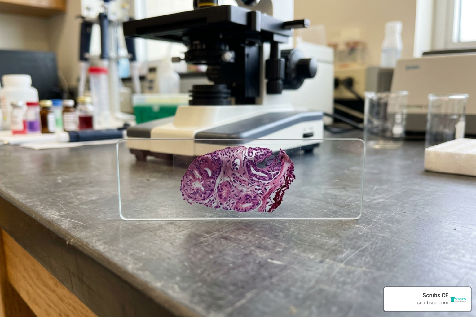
Pathological anatomy encompasses four primary subspecialties:
- Surgical Pathology: The largest subfield, involving the microscopic evaluation of tissues removed during surgical procedures (such as biopsies and organ resections) to diagnose disease and guide surgical strategy.
- Cytopathology: The study of individual cells or small clusters of cells shed naturally or scraped/aspirated from mucosal surfaces and body cavities (such as cervical Pap smears or thyroid fine-needle aspirations).
- Molecular Pathology: The examination of nucleic acids (DNA/RNA) and gene expression patterns within tissue samples to identify precise genetic mutations, oncogenic drivers, and therapeutic targets.
- Forensic Pathology: The application of pathological anatomy in legal and medicolegal settings, utilizing post-mortem autopsy techniques to determine the cause, mechanism, and manner of death.
Surgical Pathology and Biopsy Analysis
Surgical pathology represents the daily operational core of most anatomical pathology departments. Pathologists in this field evaluate biological tissue specimens collected through various clinical methods:
- Core Needle Biopsies: Small, cylindrical tissue cores extracted using specialized hollow needles, frequently used for evaluating suspicious breast, prostate, or liver lesions.
- Incisional Biopsies: Surgical sampling of a representative portion of a larger mass or lesion to establish a preliminary diagnosis before major intervention.
- Excisional Biopsies: Complete surgical removal of an entire suspicious lesion or polyp (such as a cauliflower-shaped colorectal polyp) for simultaneous diagnosis and definitive localized treatment.
- Surgical Resections: Large-scale organ resections (such as a colectomy or mastectomy) performed to remove widespread disease along with surrounding lymph nodes and healthy tissue boundaries.
During surgical pathology evaluation, gross examination is conducted first. Pathologists carefully inspect, measure, weigh, section, and ink the specimen margins. Inking tissue edges allows the pathologist to verify under the microscope whether a tumor has been excised with clear, healthy tissue margins or if tumor cells extend directly to the surgical cut edge.
In urgent scenarios, surgeons request an intraoperative frozen section consultation. The pathologist rapidly freezes fresh tissue using a cryostat microtome, cuts a section, stains it, and provides an immediate diagnosis within minutes to inform the surgeon whether margins are clear while the patient remains on the operating table.
Cytopathology and Forensic Pathology
Cytopathology offers a minimally invasive alternative to solid tissue biopsies. Rather than examining intact tissue architecture, cytopathologists evaluate individual cell morphology.
A classic example is the cervical Papanicolaou (Pap) test, introduced by George Papanicolaou in 1928, which revolutionized early screening for cervical dysplasia and carcinoma. Another common method is Fine-Needle Aspiration (FNA), where thin needles harvest cellular fluid from palpable masses in the thyroid, lymph nodes, or salivary glands. Cytotechnologists perform initial high-volume slide screening, escalating atypical or suspicious preparations to board-certified pathologists for final evaluation.
Forensic pathology applies these same diagnostic principles post-mortem. Forensic pathologists perform systematic autopsies to investigate sudden, unexpected, violent, or suspicious deaths. By carefully prosecting tissues, examining gross organ trauma, and evaluating toxicological and microscopic evidence (such as identifying gray-yellow purulent pus over the cerebral meninges in acute bacterial meningitis), forensic specialists establish legal causes of death to assist law enforcement, coroners, and public health authorities.
Core Diagnostic Techniques and Technological Advances
To transform raw biological tissue into actionable diagnostic reports, pathological anatomy relies on a sophisticated continuum of laboratory techniques. Modern laboratories combine traditional optics with advanced molecular diagnostics.
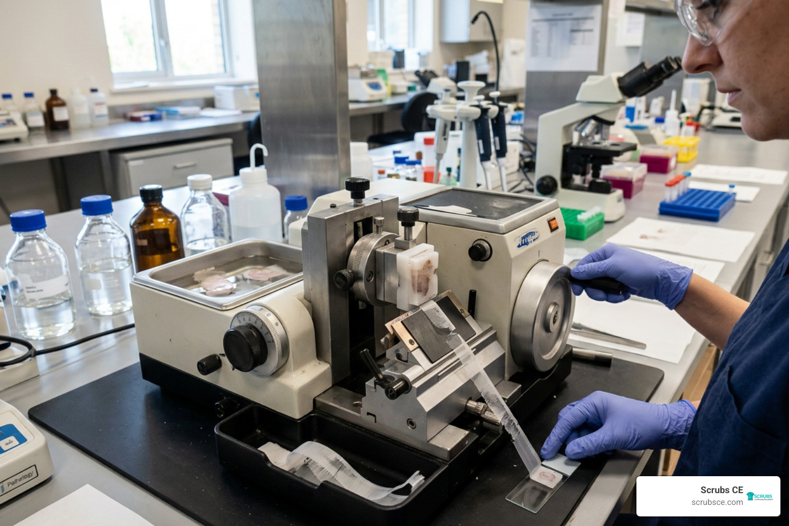
Microscopic Techniques and Standard Staining
The foundational diagnostic workhorse of pathological anatomy remains light microscopy coupled with Hematoxylin and Eosin (H&E) staining. However, before a tissue section can be viewed under a microscope, it must undergo rigorous preparation:
- Fixation: Freshly excised tissue is immersed in a preservative liquid—most commonly 10% neutral buffered formalin—to arrest autolysis, inhibit bacterial degradation, and cross-link cellular proteins.
- Processing and Paraffin Embedding: The fixed tissue is dehydrated through ascending alcohol baths, cleared in xylene, and infiltrated with molten paraffin wax. Once cooled, the tissue is encased in a solid paraffin block.
- Microtome Sectioning: A precision microtome slices the paraffin block into ultra-thin tissue sections measuring just 3 to 5 microns thick—thin enough to allow light transmission without cell overlap.
- Staining: Sections are mounted on glass slides and stained with H&E. Hematoxylin, a basic dye, binds to nucleic acids, coloring cell nuclei deep blue/purple. Eosin, an acidic dye, stains cytoplasmic proteins and extracellular collagen varying shades of pink.
For specialized diagnostic questions, pathologists utilize supplementary histochemical stains:
- Periodic Acid-Schiff (PAS): Highlights complex carbohydrates, basement membranes, and fungal cell walls.
- Ziehl-Neelsen: Detects acid-fast mycobacteria, such as Mycobacterium tuberculosis.
- Elastic Stains: Visualize disrupted vascular elastic laminae, helping confirm vasculitis (such as in granulomatous polyangiitis).
For further technical insights into tissue preparation, explore these Pathological Anatomy Techniques.
The Pathological Anatomy Definition in Diagnostic Oncology
While traditional morphology under H&E staining provides essential clues, morphologically identical tumors can behave entirely differently. Modern pathological anatomy resolves these limitations through ancillary testing—most notably Immunohistochemistry (IHC), In Situ Hybridization (ISH), and Next-Generation Sequencing (NGS).
Immunohistochemistry uses antigen-antibody specificity to tag intracellular markers with chromogenic enzymes. For example, if a metastatic tumor of unknown origin presents in a lymph node, an IHC panel testing for cytokeratins, S100, or leukocyte common antigen can immediately distinguish carcinoma from melanoma or lymphoma.
In oncology, IHC and molecular diagnostics do more than establish a diagnosis; they dictate targeted therapy:
- HER2/neu Overexpression: Identified via IHC or Fluorescence In Situ Hybridization (FISH) in invasive ductal breast carcinoma, predicting response to targeted therapies like trastuzumab.
- PD-L1 Expression: Quantified on tumor cell membranes to predict responsiveness to immune checkpoint inhibitors.
- Molecular Driver Mutations: Polymerase Chain Reaction (PCR) and NGS pinpoint specific genetic alterations (such as EGFR mutations in non-small cell lung carcinoma or BRAF V600E mutations in melanoma), allowing oncologists to prescribe tailored molecular inhibitors.
Clinical Role and Interdisciplinary Collaboration
Pathologists are frequently referred to as “the doctor’s doctor.” Although they rarely interact directly with conscious patients, their analytical findings guide virtually every major clinical decision in patient care.
Tumor Boards and Surgical Margin Evaluation
Modern oncology relies heavily on multidisciplinary collaboration. Pathologists serve as central figures on hospital tumor boards—regular consultative panels where surgeons, medical oncologists, radiation oncologists, and diagnostic imaging specialists gather to discuss complex patient cases.
During tumor board reviews, the pathologist presents tissue slides, reviews histopathological grades, clarifies staging classifications, and reports surgical margin status. If microscopic evaluation reveals tumor cells resting directly on an inked resection edge, the team knows immediate re-excision or adjuvant radiation therapy is required to prevent local recurrence.
Bridging Pathology with Diagnostic Radiology
A critical area of clinical collaboration is radiologic-pathologic correlation. Diagnostic imaging modalities—such as CT, MRI, ultrasound, and mammography—identify structural abnormalities, tissue density shifts, and space-occupying lesions deep within body cavities. However, imaging findings require structural tissue validation to confirm exact disease entities.
For radiologic technologists, radiographers, and medical imaging professionals, understanding the underlying pathological anatomy enhances clinical practice. When imaging specialists understand how pathological processes—such as calcifications, osteolytic destruction, or necrotic tumor centers—manifest anatomically, they can capture optimized diagnostic images.
Professional credentials administered by the AMERICAN REGISTRY OF RADIOLOGIC TECHNOLOGISTS® (ARRT®) emphasize the importance of understanding pathology alongside anatomical positioning. Maintaining strong continuing education standards certified through organizations like the ARRT® and the ASRT® (American Society of Radiologic Technologists) ensures that imaging teams work seamlessly with pathologists to maintain diagnostic precision.
Frequently Asked Questions About Pathological Anatomy
What is the primary difference between anatomical pathology and clinical pathology?
Anatomical pathology (or pathological anatomy) focuses on structural changes in solid tissues, organs, and cellular samples obtained via biopsy, surgery, or autopsy. Clinical pathology deals with the quantitative laboratory analysis of bodily fluids, such as blood, urine, cerebrospinal fluid, and metabolic markers.
How did Rudolf Virchow transform pathological anatomy?
In the mid-19th century, Rudolf Virchow introduced cellular pathology (Cellularpathologie). He demonstrated that disease originates within structural and functional disruptions of individual cells rather than whole-organ dysfunction or ancient fluid humors, establishing the microscopic foundation of modern diagnostic medicine.
What are the main techniques used to examine tissue samples in pathological anatomy?
The primary techniques include gross macroscopic examination, tissue fixation, paraffin embedding, microtome sectioning, standard Hematoxylin and Eosin (H&E) staining, specialized histochemical stains, Immunohistochemistry (IHC), In Situ Hybridization (ISH), and advanced molecular testing such as PCR and Next-Generation Sequencing (NGS).
Conclusion
Understanding the pathological anatomy definition reveals why this discipline remains the absolute bedrock of diagnostic medicine. By bridging macroscopic clinical observations with microscopic cellular architecture and molecular genetics, pathological anatomy allows healthcare teams to deliver definitive disease diagnoses, evaluate prognoses, and tailor targeted therapies for individual patients.
As personalized medicine, digital pathology platforms, and automated molecular diagnostics continue to evolve, the structural insights provided by pathological anatomy will remain central to patient outcomes. For healthcare professionals seeking to sharpen their anatomical knowledge and meet mandatory licensure requirements, maintaining high-quality continuing education is essential.
At Scrubs CE, we provide self-paced, affordable online continuing education designed to empower imaging professionals, radiographers, and healthcare teams. Expand your diagnostic insight today by exploring our comprehensive course on Anatomy for Radiographers!