
How to Keep Patients Safe and Calm During Nuclear Medicine Scans
Why Nuclear Medicine Radiation Safety Matters
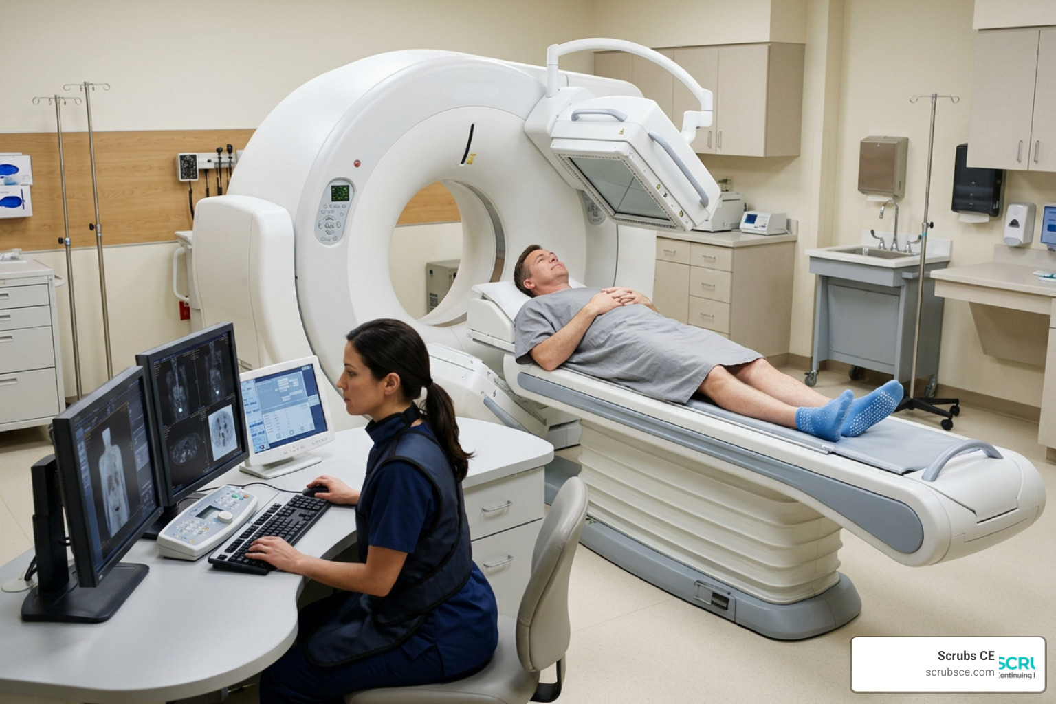
Nuclear medicine radiation safety is the vital shield that protects patients and healthcare workers while they use invisible, life-saving radioactive tools.
If you are looking for a quick reference on key safety guidelines, here is what you need to know:
| Safety Category | Key Guideline / Limit |
|---|---|
| Core Principle | ALARA (Keep doses As Low As Reasonably Achievable) |
| Occupational Worker Dose Limit | 5 rem (50 mSv) per year |
| Pregnant Worker Dose Limit | 0.5 rem (5 mSv) over the entire pregnancy |
| Patient Release Limit | Allowed if the dose rate is under 7 mrem/hr at 1 meter |
| Minor Spill Threshold | Under 100 mCi for Technetium-99m; under 1 mCi for Iodine-131 |
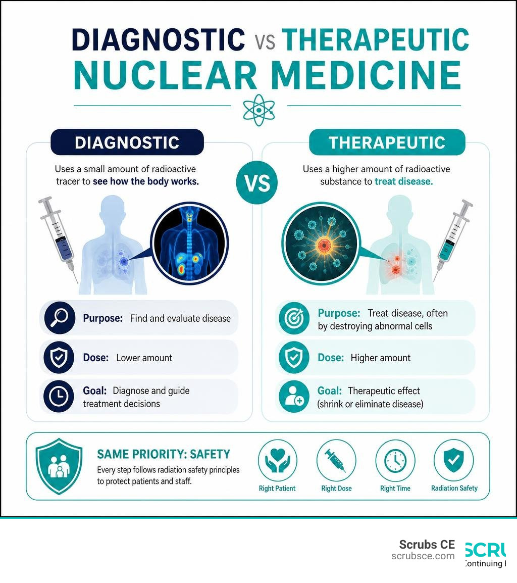
Every year, more than 20 million Americans benefit from nuclear medicine. These procedures use tiny amounts of radioactive drugs—called radiopharmaceuticals—to find and treat serious diseases. While diagnostic scans help us see how organs function, therapeutic doses actively target and destroy cancer cells.
To keep patients calm and safe, you must balance clinical precision with deep empathy.
I am Zita Ewert, founder of Scrubs CE, where I specialize in creating easy-to-understand continuing education courses on Nuclear medicine radiation safety for busy imaging professionals. Over the years, I have helped thousands of technologists satisfy their licensing requirements for the AMERICAN REGISTRY OF RADIOLOGIC TECHNOLOGISTS® (ARRT®).
Quick Nuclear medicine radiation safety definitions:
- Nuclear medicine continuing education
- Nuclear medicine technology continuing education
- Nuclear medicine technologist courses
Core Principles of Nuclear Medicine Radiation Safety
To build a strong safety culture, we must master the core principles of radiation protection. These principles keep our teams safe and reassure nervous patients that their care is managed with extreme precision.
The three pillars of radiation protection are:
- Justification: Every exposure must show a net benefit. We must ensure the medical value of the scan or therapy outweighs the small risk of radiation exposure.
- Optimization: This is where we apply the ALARA (As Low As Reasonably Achievable) principle. We use the smallest amount of a radiopharmaceutical needed to get clear, diagnostic-quality images.
- Dose Limitation: We apply strict legal dose limits to occupational workers and the general public. (Note: These limits do not apply to medical exposures for the patients themselves, as their dosages are tailored to their specific clinical needs.)
When we handle unsealed radioactive materials in our daily workflows, we rely on the TDS principle (Time, Distance, and Shielding) to minimize external exposure:
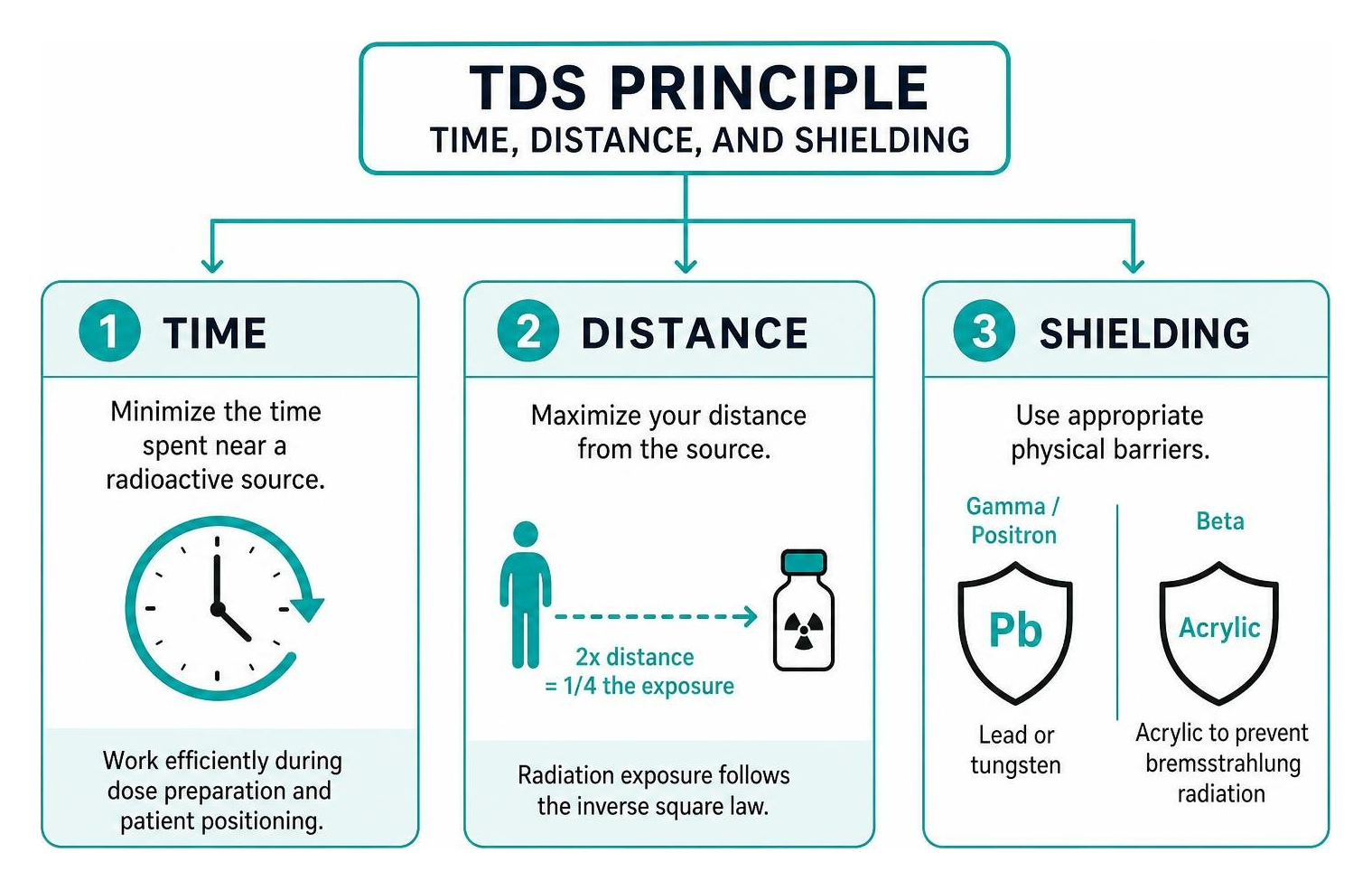
- Time: Minimize the time spent near a radioactive source. Work efficiently during dose preparation and patient positioning.
- Distance: Maximize your distance from the source. Since radiation exposure follows the inverse square law, doubling your distance from a patient or vial reduces your exposure to one-fourth of the original level.
- Shielding: Use appropriate physical barriers. Lead or tungsten shields are essential when handling gamma and positron emitters, while acrylic is preferred for beta emitters to prevent bremsstrahlung radiation.
For a deeper dive into clinical safety management, you can read the Nuclear Medicine Safety – StatPearls – NCBI Bookshelf reference.
Understanding Radiation Biology and Physics
To explain radiation safety to others, we must understand how ionizing radiation interacts with human tissue. Radiation effects are split into two main categories:
- Deterministic Effects: These are predictable, threshold-dependent physical responses. If a dose exceeds a certain limit, tissue damage occurs (such as skin reddening or hair loss). In diagnostic nuclear medicine, we rarely see these effects because our diagnostic doses are kept far below these thresholds.
- Stochastic Effects: These are probabilistic effects with no established threshold. The most notable stochastic effect is radiation-induced cancer. While the probability of a stochastic effect increases with a higher dose, the severity does not.
To stay conservative, radiation safety programs use the Linear No-Threshold (LNT) hypothesis. This model assumes that any exposure to ionizing radiation, no matter how small, carries some risk of biological damage. While some researchers debate if low-dose radiation (under 100 mSv) is truly harmful, the LNT model remains our gold standard for daily safety protocols.
We must also match our safety precautions to the specific emissions of the isotopes we use:
- Alpha Particles (e.g., Radium-223): These heavy particles have high linear energy transfer (LET) but a very short range (micrometers in tissue). They pose no external exposure hazard because they cannot penetrate the skin. However, they are highly destructive if swallowed or inhaled. Strict fluid-handling precautions are vital.
- Beta Particles (e.g., Lutetium-177, Yttrium-90): These particles travel a few millimeters to centimeters in tissue. They present a localized hazard to skin and eyes. We shield them with low-density materials like acrylic to avoid generating secondary X-rays.
- Gamma Photons (e.g., Technetium-99m, Iodine-131): These highly penetrating electromagnetic waves can escape the patient’s body entirely. They travel long distances and require dense lead or tungsten shielding to protect staff and nearby patients.
To master these concepts and earn your required CEUs, check out our course on Stay Safe Stay Certified Mastering Radiation Protection CEUs.
Implementing Nuclear Medicine Radiation Safety in Daily Practice
Putting safety principles into action requires specialized equipment and strict quality control. In the hot lab, unshielded materials must never be handled directly. We use tungsten or lead syringe shields, vial shields, and remote handling tools like forceps to keep our fingers safe from high extremity doses.
To track our personal exposure, we wear personal dosimetry badges (such as thermoluminescent dosimeters or optically stimulated luminescence badges) on our collars. We also wear ring badges on our dominant hands to monitor extremity exposure during dose preparation.
Our daily quality control routine relies heavily on the dose calibrator. Under federal guidelines, dose calibrators must maintain an accuracy of ±10% to ensure patients receive the exact prescribed amount of radioactivity.
Furthermore, the administered dosage must not deviate from the prescribed dosage by more than 20% without authorization from an Authorized User (AU). If a dosage falls outside this range, it may trigger a reportable medical event.
If you practice in the Golden State, make sure you stay compliant with local regulations by reviewing our guide on California Radiation Safety.
Regulatory Standards and Personnel Roles
Nuclear medicine is highly regulated to protect workers, patients, and the public. In the United States, the Nuclear Regulatory Commission (NRC) sets the legal standards under Title 10 of the Code of Federal Regulations (specifically 10 CFR Part 35).
Globally, organizations like the International Commission on Radiological Protection (ICRP) and the International Atomic Energy Agency (IAEA) establish international benchmarks. They publish detailed safety guides, which you can explore on the Human Health Campus – Radiation protection of workers and the public in nuclear medicine platform.
Key Personnel: AU, RSO, and RSC
Maintaining a safe department requires a clear chain of responsibility:
- Authorized User (AU): This is a physician who has met strict training requirements (such as 700 hours of specialized training for diagnostic pathways or 80 hours of classroom work for therapeutic pathways). The AU is legally responsible for prescribing and directing the administration of radiopharmaceuticals. They must sign a written directive for any therapeutic administration, or for any diagnostic dose of Sodium Iodide Iodine-131 exceeding 30 microcuries (μCi).
- Radiation Safety Officer (RSO): The RSO manages the daily operations of our radiation protection program. They ensure regulatory compliance, investigate overexposures, perform routine area surveys, and coordinate emergency responses to spills or accidents.
- Radiation Safety Committee (RSC): Required at larger medical centers, the RSC includes the RSO, an AU, a representative from nursing, and a representative from management. The committee meets regularly to review safety programs, audit exposure records, and establish local policies.
Established Dose Limits for Workers and the Public
To ensure safety, we must adhere strictly to established annual dose limits. The table below outlines these regulatory thresholds:
| Exposed Group | Annual Effective Dose Limit | Equivalent Dose Limit to Lens of Eye | Equivalent Dose Limit to Skin/Extremities |
|---|---|---|---|
| Occupational Workers | 5 rem (50 mSv) | 15 rem (150 mSv) | 50 rem (500 mSv) |
| Pregnant Workers (Fetal Limit) | 0.5 rem (5 mSv) for entire gestation | N/A | N/A |
| General Public | 0.1 rem (1 mSv) | N/A | N/A |
For pregnant workers, declaring a pregnancy is voluntary. Once declared in writing, the lower limit of 0.5 rem (5 mSv) applies to the fetus to protect it during sensitive stages of development.
Clinical Protocols: Spills, Waste, and Patient Release
Working with unsealed liquid radionuclides means we must be prepared for accidental contamination and manage our waste streams responsibly.
Managing Major vs. Minor Radioactive Spills
If a spill occurs, we must act quickly. Spills are classified as major or minor based on the radionuclide involved and its activity level:
- Technetium-99m (Tc-99m) and Thallium-201 (Tl-201): The major spill threshold is 100 mCi.
- Indium-111 (In-111) and Gallium-67 (Ga-67): The major spill threshold is 10 mCi.
- Iodine-131 (I-131): Because of its volatility and long half-life, the major spill threshold is just 1 mCi.
For a minor spill, the local staff can clean it up:
- Confine: Cover the spill with absorbent paper to prevent it from spreading.
- Clear: Warn others and clear the immediate area.
- Clean: Clean the spill using protective gloves and disposable materials, working from the outside of the spill inward.
- Survey: Use a Geiger-Mueller survey meter to check the area and personnel for remaining contamination.
- Report: Document the incident and notify the RSO.
For a major spill, we must escalate the response immediately:
- Clear: Evacuate the room and close the door to prevent access.
- Shield: If possible, place a lead shield over the spill, but do not attempt to clean it up yourself.
- Notify: Contact the RSO immediately.
- Decontaminate: Remove contaminated clothing and wash affected skin with mild soap and lukewarm water (never scrub harshly, as this can break the skin barrier).
Patient Release Criteria and Post-Therapy Guidelines
After receiving therapeutic radiopharmaceuticals, patients temporarily emit radiation. Under NRC regulations, we can release a patient if the total dose to any individual coming into contact with them is unlikely to exceed 5 mSv (0.5 rem).
To simplify this, the NRC allows patient release without complex calculations if:
- The administered activity of Iodine-131 is less than 33 mCi, or
- The measured dose rate is under 7 mrem/hr (0.07 mSv/hr) at 1 meter.
When a patient is released, we must provide them with clear, written instructions on how to minimize exposure to family members and the public. These instructions include:
- Sleeping in a separate bed.
- Using a dedicated bathroom and flushing twice.
- Avoiding close contact with children and pregnant women.
We must also warn patients about travel security. Highly sensitive radiation monitors at airports and federal buildings can detect residual radiation for weeks or months.
Here is how long common isotopes may remain detectable:
- Fluorine-18 (F-18): Typically undetectable after 1 day.
- Technetium-99m (Tc-99m): Typically undetectable after 3 to 4 days.
- Thallium-201 (Tl-201): Can remain detectable for up to 30 days.
- Lutetium-177 (Lu-177): Can remain detectable for up to 2 months.
- Iodine-131 (I-131): Can remain detectable for up to 3 months.
We always recommend providing patients with a signed travel letter explaining their treatment to prevent unnecessary delays at security checkpoints.
Patient-Centered Care and Communication Strategies
The word “radiation” can cause immediate anxiety for patients. As technologists, our job is to demystify the procedure and help them feel safe.

When talking to patients, we should use clear, reassuring language:
- Avoid jargon: Instead of saying “we are injecting an open-source radionuclide,” say “we are administering a small amount of a radioactive tracer that acts like a temporary highlighter for your cells.”
- Put risks in perspective: Explain that the radiation dose from a diagnostic scan is similar to what they naturally absorb from the environment over a few months.
- Highlight the benefits: Emphasize that nuclear medicine shows how their body is functioning in real-time, helping their doctor make accurate treatment decisions without the need for invasive procedures.
For example, when performing a cardiac study, explaining the process clearly can significantly lower a patient’s heart rate and anxiety. You can read more about these clinical applications in our guide on Nuclear Medicine Myocardial Perfusion.
Vulnerable Populations and Nuclear Medicine Radiation Safety
We must take extra precautions when imaging children, pregnant patients, or breastfeeding mothers:
- Pediatric Patients: Children are more sensitive to radiation and have a longer lifetime to develop potential stochastic effects. We always use weight-based dosing guidelines (such as the North American Consensus Guidelines) to minimize their exposure while maintaining diagnostic quality.
- Pregnancy: We must screen all female patients of childbearing age for pregnancy. If a pregnant patient requires a scan, we must carefully justify the procedure. If it is medically necessary, the clinical team will optimize the dose and scan parameters to minimize fetal exposure.
- Breastfeeding Mothers: Many radiopharmaceuticals can pass into breast milk. We must coordinate with the patient to plan temporary breastfeeding interruption times:
- Tc-99m-pertechnetate: Interrupt breastfeeding for 12 hours.
- In-111-octreotide: Interrupt breastfeeding for 60 hours (2.5 days).
- I-131-sodium iodide: Requires complete cessation of breastfeeding for that child, as the isotope can damage the infant’s thyroid gland.
For more information on handling diverse imaging cases, see our article on Infection and Inflammation in Nuclear Medicine Imaging.
Bridging the Information Gap for Outside Healthcare Professionals
A common challenge in hospitals is the communication gap between nuclear medicine specialists and non-specialist healthcare professionals (HCPs). If a patient who was recently treated with a therapeutic isotope (such as Lutetium-177) visits an outside clinic or emergency department, it can cause unnecessary panic among staff.
To prevent this, we recommend a dual-alert communication system:
- EMR Flag: An automatic alert in the Electronic Medical Record that triggers upon radiopharmaceutical administration and expires after 7 days.
- Physical Wristband: A waterproof, color-coded wristband worn by the patient. The wristband should list the radionuclide, the administration date, and a QR code linking directly to safety instructions for outside HCPs.
This simple system ensures that nurses and physicians outside our department know they can safely provide routine care (like taking blood pressure or conducting a brief consultation) by maintaining a reasonable distance and wearing standard personal protective equipment (PPE).
Frequently Asked Questions about Nuclear Medicine Safety
How does nuclear medicine radiation compare to natural background radiation?
The typical American absorbs about 6 mSv of radiation annually. Roughly 50% comes from natural background sources (like radon gas, cosmic rays, and naturally occurring radioactive materials in our bodies), while about 40% comes from medical radiation.
A standard diagnostic nuclear medicine scan (such as a bone scan or a hepatobiliary scan) typically delivers a dose of 3 to 6 mSv, which is comparable to a few years of natural background exposure.
What constitutes a reportable medical event in nuclear medicine?
Under NRC regulations, a reportable medical event occurs when a radiopharmaceutical administration is given to the wrong patient, is the wrong drug, uses the wrong route of administration, or the administered dosage deviates by more than 20% from the prescribed dosage, AND results in:
- A total effective dose equivalent (TEDE) exceeding 5 rem (50 mSv), or
- A single-organ committed dose equivalent exceeding 50 rem (500 mSv).
If a medical event occurs, we must notify the NRC by phone within 1 day, submit a written report within 15 days, and notify the referring physician and the patient within 24 hours.
Why do patients need a travel letter after radiopharmaceutical therapy?
Highly sensitive radiation portal monitors at border crossings, airports, and federal buildings can detect even trace amounts of radiation.
Because therapeutic isotopes like Iodine-131 can remain detectable for up to 3 months, a signed letter from an Authorized User prevents security delays by verifying that the radiation source is medical and safe.
Conclusion
Creating a strong safety culture in nuclear medicine is a team effort. By combining strict technical standards with clear, empathetic communication, we can protect our staff, comply with regulatory bodies, and help our patients feel safe and calm during their procedures.
If you are an imaging professional looking to stay certified and expand your clinical knowledge, we are here to help. At Scrubs CE, we offer convenient, self-paced, and affordable online continuing education courses.
Our high-quality courses provide instant certificates to help you meet your state licensing and AMERICAN REGISTRY OF RADIOLOGIC TECHNOLOGISTS® (ARRT®) requirements.
Ready to earn your next CE credits? Explore our complete catalog of Nuclear Medicine CE courses today!