
Essential PPE Requirements Every CT Tech Needs on the Job
Why CT Tech PPE Requirements Matter Every Shift

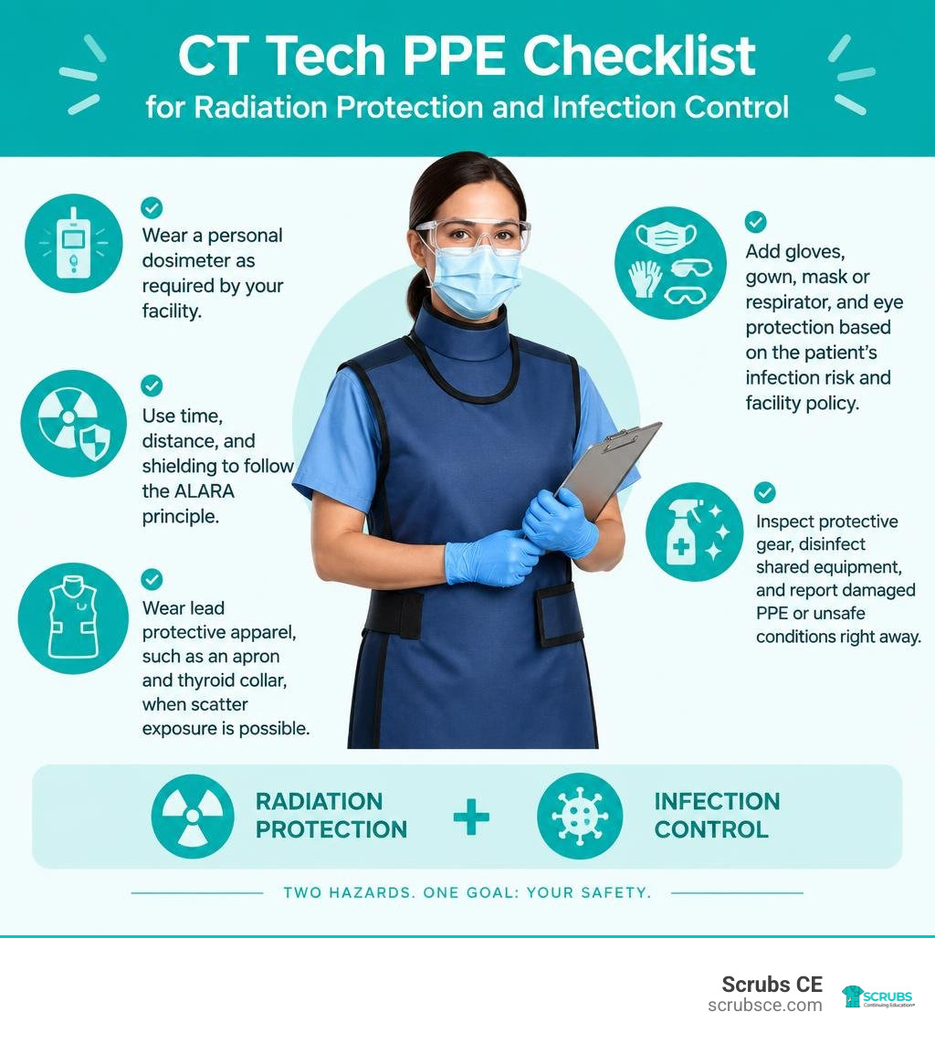
CT tech PPE requirements combine radiation shielding and infection control. In practical terms, every technologist should:
- Wear a personal dosimeter as required by the facility.
- Use time, distance, and shielding to follow the ALARA principle.
- Wear lead protective apparel, such as an apron and thyroid collar, when scatter exposure is possible.
- Add gloves, gown, mask or respirator, and eye protection based on the patient’s infection risk and facility policy.
- Inspect protective gear, disinfect shared equipment, and report damaged PPE or unsafe conditions right away.
The exact combination changes by task. A technologist operating the CT console behind a fixed barrier has a different exposure risk than someone assisting a patient, performing portable imaging, or supporting an interventional CT procedure. Good PPE protects staff from two workplace hazards at once: ionizing radiation and infectious materials.
I am Zita Ewert, a continuing-education leader for imaging professionals and the driving force behind SCRUBS Continuing Education®. My work supporting accessible, accredited education for radiologic professionals includes practical guidance on CT tech PPE requirements, radiation safety, and safe clinical practice.
Ct tech ppe requirements terms to learn:
Core Radiation Shielding and CT Tech PPE Requirements
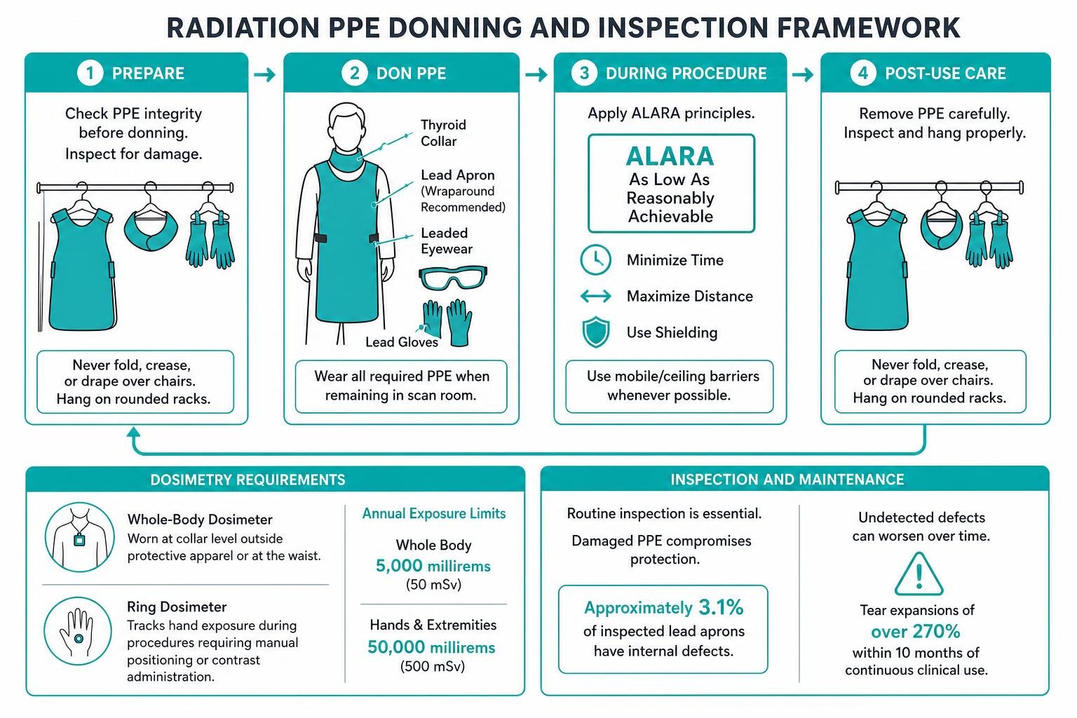
Working with computed tomography means balancing the diagnostic power of ionizing radiation with rigorous defense mechanisms. Ionizing radiation is an invisible hazard; while we cannot see or feel scatter radiation during a scan, cumulative exposure poses documented biological risks over a career. To manage this safely, the industry follows the ALARA principle: keeping exposure As Low As Reasonably Achievable by managing time, maximizing distance, and utilizing shielding.
Occupational safety standards set strict annual radiation exposure limits: 5,000 millirems (50 mSv) per year for the whole body and 50,000 millirems (500 mSv) annually for the hands and extremities. To track these doses, technologists must wear personal dosimeter badges consistently. Whole-body dosimeters are typically worn at the collar level outside protective apparel or at the waist, while ring dosimeters track hand exposure during procedures requiring manual positioning or contrast administration. Deepening your understanding through our course on radiation protection in medical radiography reinforces how these regulatory limits translate to clinical practice.
The table below outlines common protective apparel materials and their shielding characteristics:
| PPE Type | Standard Thickness (Lead Equiv.) | Scatter Attenuation Rate | Primary Protection Focus | Material Options |
|---|---|---|---|---|
| Frontal / Wraparound Apron | 0.25 mm – 0.5 mm | 90% – 95%+ | Torso, gonads, vital organs | Traditional lead, lead-free alloys (bismuth, tungsten) |
| Thyroid Collar | 0.25 mm – 0.5 mm | Up to 95% | Thyroid gland | Lightweight composite, lead-lined |
| Leaded Eyewear | 0.5 mm – 0.75 mm | 85% – 90%+ | Eye lens (cataract prevention) | Lead glass, polycarbonate leaded lenses |
| Protective Gloves | 0.25 mm – 0.5 mm | 75% – 85% | Hands and fingers | Attenuating leaded rubber / vinyl |
| Mobile / Ceiling Barriers | 0.5 mm – 1.0 mm | 99%+ | Full-body scatter deflection | Leaded acrylic, mobile lead shields |
Essential Radiation Shielding Equipment
Whenever a technologist must remain inside the scan room while the X-ray tube is energized—such as during CT fluoroscopy, interventional biopsies, or when stabilizing pediatric patients—full radiation PPE is mandatory.
- Lead Aprons and Thyroid Collars: A standard apron attenuates scatter radiation by 90% or more. Pairing a full-body apron with a fitted thyroid collar boasting at least 0.25 mm—and preferably 0.5 mm—lead equivalence reduces scatter radiation exposure to radiosensitive neck tissues by up to 95%. Wraparound aprons are strongly recommended in interventional settings where clinicians frequently turn their backs to the gantry.
- Leaded Eyewear: Radiation-induced cataracts represent a significant occupational hazard from chronic low-dose scatter. Wearing leaded glasses with side shields attenuates significant scatter and preserves ocular health over decades of practice.
- Protective Lead Gloves and Barriers: While lead gloves reduce tactile sensitivity during fine needle placement, specialized attenuating exam gloves offer a practical balance between protection and manual dexterity. When possible, mobile leaded acrylic barriers should be positioned between the technologist and the gantry to deflect primary scatter.
Maintenance, Inspection, and Defect Detection
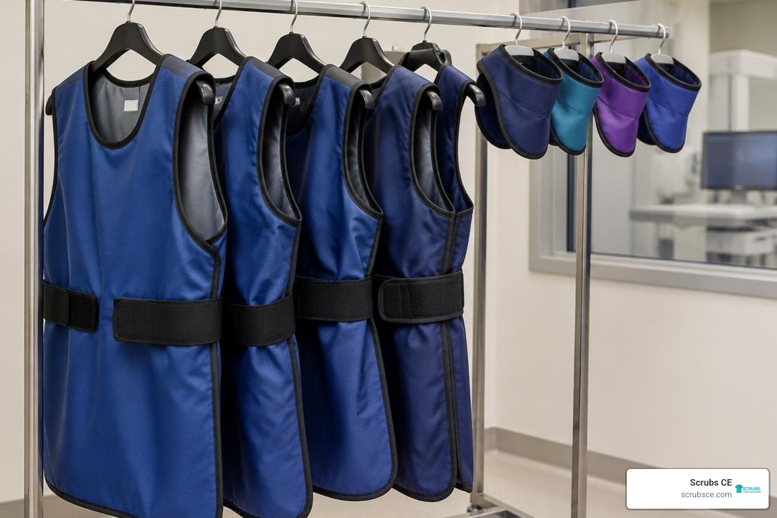
Personal radiation shielding only works if the integrity of the attenuating core remains intact. Protective garments must never be folded, creased, or draped over chairs, as folding breaks the internal lead lining. Always hang aprons and collars on heavy-duty, rounded racks specifically designed for radiation gear.
Routine maintenance protocols are vital. Clinical studies have revealed that approximately 3.1% of inspected lead aprons harbor internal defects that allow unsafe radiation transmission. More critically, aprons with small undetected defects can experience tear expansions of over 270% within 10 months of continuous clinical use.
Every facility must establish a documented, annual visual and fluoroscopic inspection program. Technologists should inspect garments under low-dose fluoroscopy or radiography to identify hidden cracks, thinning, or internal tears. Any garment demonstrating significant holes, cracks near critical organs, or severe seam separation must be taken out of service immediately and disposed of according to hazardous waste regulations. Technologists can refresh their compliance skills through dedicated radiation protection CEUs to keep inspection workflows up to date.
Infection Control and Biosafety Protocols in the CT Suite
CT technologists operate at the intersection of advanced imaging and acute infection control. Because the CT suite receives patients from emergency departments, intensive care units, and outpatient clinics, rigorous biosafety protocols are necessary to prevent healthcare-associated infections.
During infectious outbreaks, CT imaging frequently plays a key diagnostic role. Diagnostic data highlighted that chest CT demonstrates exceptional sensitivity for detecting pulmonary viral patterns early, particularly when initial RT-PCR throat swab tests yielded positive detection rates of only 30% to 60% upon initial presentation (Zhao et al., 2020). To handle high clinical volumes without compromising staff safety, departments implement Biosafety Level 2 (BSL-2) or modified contact/droplet precautions.
Core infection control PPE in the CT suite includes:
- N95 or PAPR Respirators: Essential for airborne precautions and high-risk aerosol-generating environments.
- Fluid-Resistant Isolation Gowns: Impervious gowns protect skin and scrubs from infectious fluids and contrast material.
- Nitrile Gloves: Double gloving is standard practice when handling high-risk patients or during deep decontamination cycles.
- Eye Protection and Face Shields: Full-face shields prevent mucosal splash contamination during intravenous access and patient transfer.
- Disposable Booties and Head Covers: Prevent carrying pathogen reservoirs across different hospital units.
Strict Donning and Doffing Procedures for CT Tech PPE Requirements
Preventing self-contamination requires adhering to a strict, structured sequence when putting on (donning) and taking off (doffing) personal protective gear. For high-level biosafety workflows, the complete donning protocol can take up to 30 minutes to ensure every seal and overlap is secure.
Donning Sequence:
- Perform thorough hand hygiene using an alcohol-based rub or soap and water.
- Put on disposable shoe covers and hair cap.
- Don the N95 respirator, performing a seal check (inhale and exhale to ensure no air leaks around the bridge or chin).
- Put on protective eyewear or a full-face shield.
- Don the fluid-impervious gown, tying all fasteners securely at the neck and waist.
- Put on nitrile gloves, pulling the cuffs over the sleeves of the gown to create a closed seal.
Doffing Sequence (Buffer Room Workflow):
- Perform hand hygiene on gloved hands before beginning removal.
- Remove outer gloves and gown together in a rolling motion, turning the contaminated exterior inward, and dispose of them in a designated biohazard receptacle.
- Perform hand hygiene.
- Remove face shield or goggles by grasping the back strap without touching the front surface.
- Remove shoe covers and cap, followed by another round of hand hygiene.
- Remove the respirator by pulling the bottom strap over the head, followed by the top strap, avoiding contact with the filter face.
- Perform final, rigorous hand hygiene before exiting the clean area.
CT Scanner Disinfection and Environmental Modifications
Radiology departments optimize safety by implementing physical layout modifications, such as the “three areas and two passages” system (Zhao et al., 2020). This architectural workflow divides the department into:
- Contaminated Zone: The CT scanner room where the patient is positioned and imaged.
- Potentially Contaminated Zone: The control room and intermediate buffer corridors.
- Clean Zone: Technologist staff break rooms, clean supply storage, and report areas.
Buffer rooms connecting these zones allow staff to perform controlled doffing and intermediate disinfection.
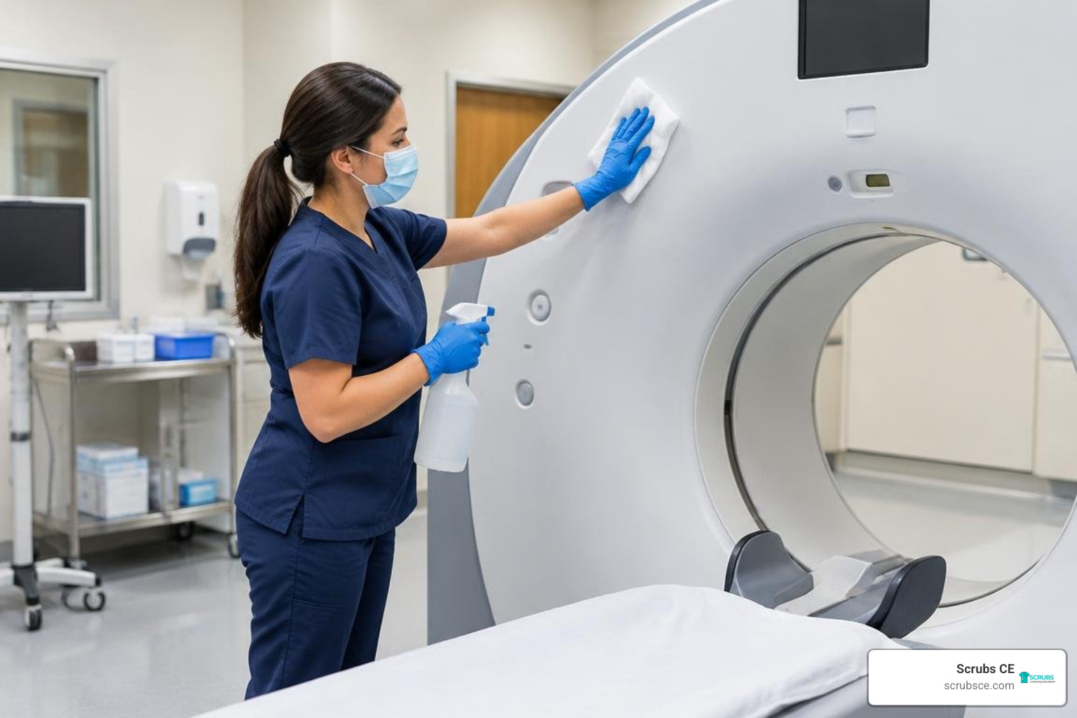
Between every scanned patient, all contact surfaces—including the patient table, head holders, straps, injector controls, and gantry console—must be thoroughly wiped down with hospital-grade disinfectants, such as 75% ethanol or quaternary ammonium solutions. For high-risk aerosol or droplet pathogens, suites should utilize ultraviolet (UV) air disinfection lamps for at least 60 minutes 2 to 3 times daily, paired with high-efficiency particulate air (HEPA) filtration cycles (Zhao et al., 2020).
Operational Safety: Workflows, Ergonomics, and Emergency Procedures
Maintaining a safe imaging environment requires looking beyond physical apparel to examine operational structures, ergonomics, and emergency mechanics. When imaging workloads are intense—such as a single technologist managing 40 to 50 CT scans during a single shift (Zhao et al., 2020)—fatigue can compromise both diagnostic accuracy and personal safety.
To prevent burnout and lapses in safety protocols, imaging departments benefit from structuring work shifts into 5- to 6-hour high-intensity rotations with scheduled rest intervals (Zhao et al., 2020).
Physical wellbeing also hinges on ergonomic workstation habits:
- Adjust control room chairs so your eyes align naturally with the top third of the monitor screens.
- Keep feet flat on the floor or supported by an ergonomic footrest.
- Use vertical ergonomic mice and keyboard wrist rests to prevent repetitive strain injuries during image reconstruction.
- Utilize mechanical sliding boards and ceiling-mounted patient transfer hoists when moving non-ambulatory patients onto the CT table.
Technologists can review essential career benchmarks by exploring comprehensive CT technologist requirements to understand modern professional expectations.
Managing Pregnant Patients and Special Populations
Imaging pregnant or potentially pregnant patients requires adherence to the ALARA framework. While ultrasound or non-ionizing modalities are preferred first-line choices, emergency diagnostic scenarios (such as suspected pulmonary embolism or major trauma) often make CT unavoidable.
In these instances, technologists should apply customized low-dose protocols, utilize automated tube current modulation (mAs), and position lead shielding over the inferior aspects of the abdomen and pelvis if the scan volume permits. Clear pre-scan screening and verification protocols prevent accidental fetal exposure, and contrast administration should always be weighed against maternal renal status and fetal thyroid considerations. For deeper clinical study, our online radiation protection and patient care course provides practical frameworks for handling sensitive patient populations.
Equipment Safety Features and Emergency Stop Protocols
Modern CT scanners are powerful electromechanical systems with gantries containing high-speed rotating X-ray tubes and heavy detector arrays. Technologists must know the exact location of emergency shut-off controls:
- Gantry Emergency Stop Buttons: Located on both the front and rear of the gantry casing. Pressing this immediately cuts electrical power to the rotating tube and mechanical drive systems, halting rotation instantly.
- Console Emergency Stop: Positioned directly on the operator console keyboard or touch panel to abort scans if a patient experiences contrast extravasation, severe allergic reactions, or unexpected movement.
- Wall Power Kill Switches: Main electrical disconnect breakers located inside the scan room and control booth.
These safety switches must be visually verified during daily pre-shift inspections and reported immediately if mechanical resistance or electrical faults appear.
Regulatory Compliance and Professional Standards for CT Technologists
Staying compliant with safety standards is a mandatory condition of professional practice. The AMERICAN REGISTRY OF RADIOLOGIC TECHNOLOGISTS® (ARRT®) and the American Society of Radiologic Technologists (ASRT®) define clinical standards that govern radiation safety, patient advocacy, and ethical conduct.
State regulatory bodies reinforce these national frameworks. For example, the Connecticut Radiographer Licensure Statutes establish formal licensure mandates, credentialing prerequisites, and legal scopes of practice for diagnostic imaging professionals. Similarly, administrative frameworks such as the State CT Operator Qualifications and Safety Guidelines outline specific requirements for equipment operation, supervised on-the-job training parameters, and radiation physics competency.
Technologists looking for comprehensive overviews can consult our computed tomography CE guide to stay ahead of evolving state and national credentialing benchmarks.
State Licensure and CT Tech PPE Requirements for Clinical Practice
Maintaining active clinical status requires recurring continuing education. Many state licensing boards mandate that radiographers complete at least 24 contact hours of approved continuing education every two years, covering radiation physics, PPE protocols, quality assurance, and infection control. Additionally, staying current with CT CEU credits ensures imaging professionals maintain their postprimary certifications with the AMERICAN REGISTRY OF RADIOLOGIC TECHNOLOGISTS® (ARRT®) while mastering contemporary imaging technologies. For a broader perspective on keeping your credentials active across multiple disciplines, check out our Radiology CE courses guide and our overview of radiologic technologist continuing education credits.
Frequently Asked Questions About CT Tech Safety and PPE
What is the annual occupational radiation exposure limit for CT technologists?
The regulatory occupational whole-body dose limit set by the Nuclear Regulatory Commission (NRC) and state radiation agencies is 5,000 millirems (50 mSv) per year. The extremity limit for hands and feet is 50,000 millirems (500 mSv) annually. Dosimeter badges track these numbers monthly or quarterly to ensure doses remain well below these thresholds.
How often should lead aprons and radiation PPE be inspected for damage?
Radiation PPE must undergo formal inspection at least once per year. This includes both a tactile/visual examination for outer cover tears and seam fraying, along with an X-ray or fluoroscopic inspection to check for internal cracks, thinning, or lead separation. Documented inspection records must be maintained for accreditation and regulatory audits.
Can lead-free radiation protective apparel provide equivalent protection to traditional lead?
Yes. Modern lead-free aprons utilize proprietary composite alloys—such as bismuth, tungsten, and antimony—to achieve radiation attenuation equivalent to 0.25 mm or 0.5 mm lead. These garments can reduce overall garment weight by up to 20% to 30%, decreasing musculoskeletal fatigue while maintaining safety compliance.
Conclusion
Personal protective equipment is the backbone of safe clinical practice in computed tomography. By pairing proper radiation shielding—such as certified lead aprons, thyroid collars, and protective eyewear—with robust infection control measures, technologists protect their long-term health while delivering high-quality diagnostic care.
Staying compliant with safety standards and ARRT® requirements is an ongoing professional commitment. At Scrubs CE, we provide self-paced, high-quality courses designed to help you meet licensure mandates, refresh your safety expertise, and advance your clinical career on your schedule. Ready to earn your mandatory credits? Explore our complete catalog of computed tomography certification and CE courses today!
References
Zhao Y, Xiang C, Wang S, Peng C, Zou Q, Hu J. “Radiology department strategies to protect radiologic technologists against COVID19: Experience from Wuhan..” European Journal of Radiology, 2020. PMCID PMC7194837.