
A Beginner Crash Course in Human Lymphatic Anatomy and Function
Anatomy and Physiology for Dummies Lymphatic System Essentials: Structure and Fluid Dynamics
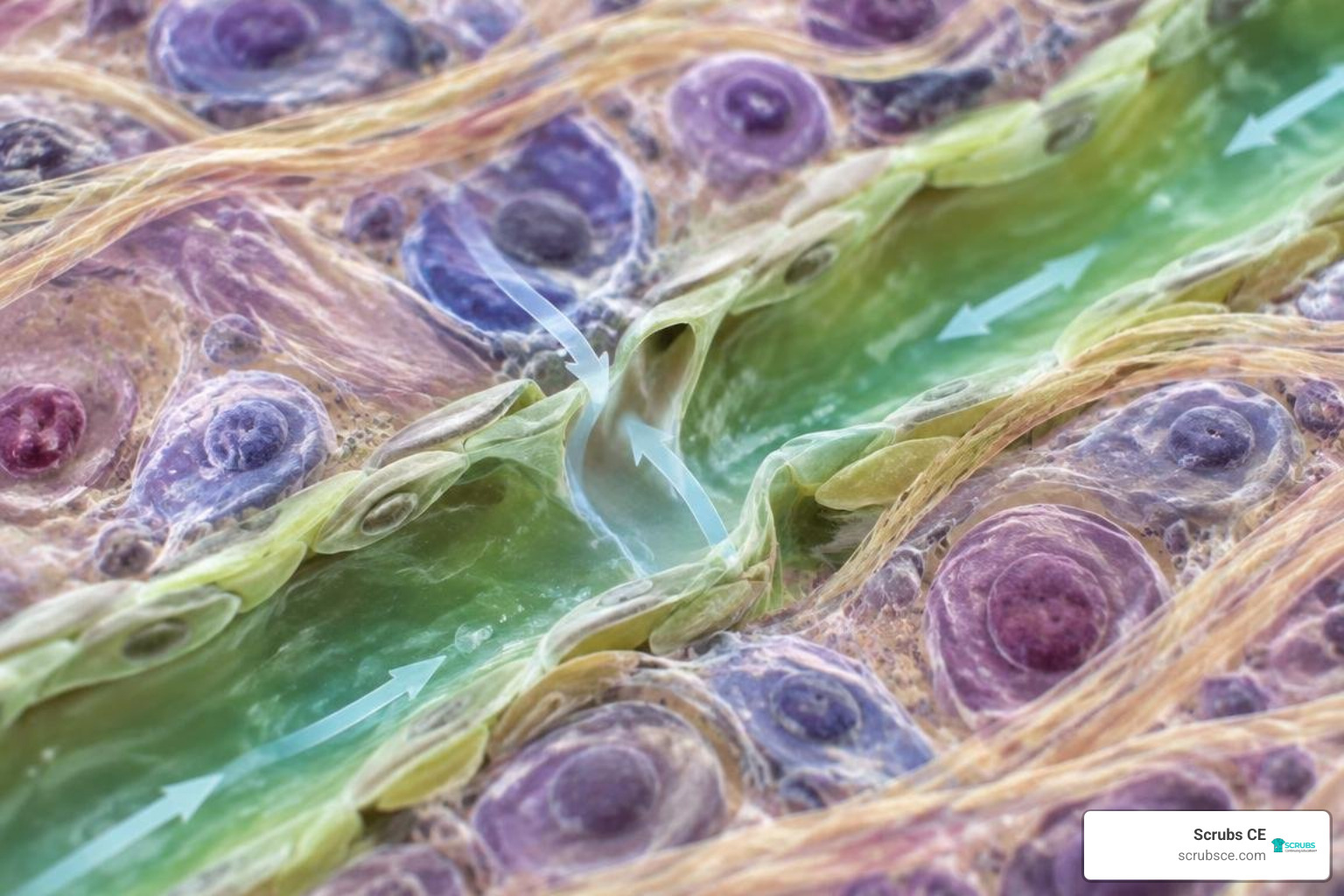
The lymphatic system is a vital vascular and immunological network responsible for maintaining fluid balance, absorbing dietary fats, and orchestrating immune defense throughout the body. As blood surges through microscopic capillaries, plasma continuously filters into surrounding interstitial spaces. The lymphatic system recovers this fluid (lymph), filters it through specialized lymph nodes, and returns it to the bloodstream to prevent tissue swelling and maintain blood volume. When reviewing anatomy and physiology for dummies lymphatic system concepts, learners explore how this indispensable partner to the circulatory system protects tissues and preserves fluid homeostasis, as highlighted in the OpenStax guide on lymphatic and immune anatomy.
Understanding Anatomy and Physiology for Dummies Lymphatic System Functions
To appreciate how this system keeps us alive, we can break its workload down into three fundamental responsibilities:
- Fluid Recovery and Balance: The primary mechanical role is returning fluid to the bloodstream. As blood moves through systemic capillaries, high hydrostatic pressure pushes clear, protein-poor plasma out into the spaces between cells (the interstitial space). The lymphatic network recovers this leftover fluid, now termed lymph, preventing severe tissue swelling (edema).
- Dietary Lipid Absorption: In the small intestine, specialized lymphatic capillaries called lacteals absorb dietary fats and fat-soluble vitamins (A, D, E, and K) that are too large to enter bloodstream capillaries directly. This milky, lipid-rich fluid is known as chyle.
- Immune Surveillance and Defense: The lymphatic network acts as the tactical highway for our immune system. It filters fluids through specialized checkpoints, traps foreign invaders, and houses billions of lymphocytes ready to launch targeted counterattacks. Developing a solid foundation in understanding anatomy & physiology helps healthcare learners visualize how these defense processes occur across multiple body systems.
How Lymph Fluid Forms and Travels Through the Body
Fluid dynamics in the human body operate on sheer scale. Every day, approximately 20 liters (about 50 pints) of plasma filters out of arterial capillaries into interstitial spaces. Of that total volume, blood capillaries reabsorb roughly 17 liters directly on the venous side.
That leaves an essential 3 liters of interstitial fluid stranded in tissue spaces. The total volume of interstitial fluid residing across the entire body at any given moment sits around 1 to 2 liters (2 to 4 pints). The lymphatic system absorbs and cleanses those leftover 3 liters daily before returning them to circulation.
Lymphatic capillaries are microscopic, blind-ended tubes woven throughout tissue spaces. Their walls consist of loosely overlapping endothelial cells that act like one-way swing doors (minivalves):
- When interstitial fluid pressure rises, these minivalves swing inward, allowing fluid, proteins, cellular debris, and pathogens to rush inside.
- Once inside, internal pressure increases, pushing the endothelial flaps shut so the collected lymph cannot escape back into the tissue.
Because the lymphatic system lacks a central mechanical pump like the heart, it relies on passive mechanical forces and pressure gradients to keep fluid moving in one direction:
- Skeletal Muscle Pump: Everyday movements contract skeletal muscles, squeezing nearby lymph vessels and propelling fluid forward.
- Respiratory Pump: Inhaling creates negative pressure inside the chest cavity, drawing lymph upward toward the thoracic region.
- Pulsations of Adjacent Arteries: Rhythmic pulses from nearby arteries compress surrounding lymphatic channels.
- Smooth Muscle Contraction: Larger lymphatic vessels contain intrinsic smooth muscle within their walls that rhythmically contracts.
- Internal One-Way Valves: Similar to veins, lymphatic vessels are packed with delicate valves that prevent backflow.

These vessels merge into larger trunks and eventually empty into two major channels:
- Right Lymphatic Duct: Drains lymph exclusively from the upper right quadrant of the body (right side of the head, neck, right thorax, and right arm) and empties into the right subclavian vein.
- Thoracic Duct: A much larger vessel that begins near the lumbar region as the cisterna chyli. It drains lymph from the entire rest of the body (both legs, abdomen, left thorax, left arm, and left side of the head) and empties into the left subclavian vein.
Primary vs. Secondary Lymphoid Organs: The Body’s Defense Stations
Lymphoid organs provide dedicated anatomical zones where immune cells generate, mature, and engage hostile foreign antigens, as detailed in the Cleveland Clinic clinical overview of the lymphatic system.
Mastering Anatomy and Physiology for Dummies Lymphatic System Organs
We categorize lymphoid structures into primary and secondary organs based on their functional roles:
Primary Lymphoid Organs (Where Immune Cells Are Born and Educated)
Primary lymphoid organs provide the specialized environments where lymphocytes originate and achieve immunocompetence (the ability to recognize specific antigens without attacking self-tissues).
- Red Bone Marrow: The soft, vascular tissue found inside the spongy sections of bones (such as the pelvis, sternum, and femur heads). It houses hematopoietic stem cells that give rise to all blood cells. B lymphocytes (B cells) both originate and complete their maturation here.
- Thymus Gland: Located in the superior mediastinum behind the sternum, the thymus serves as the training school for T lymphocytes (T cells), which migrate from the bone marrow to mature and undergo rigorous self-tolerance screening. The thymus is prominent in infants and children. Following puberty, it undergoes thymic involution, shrinking at a rate of approximately 3% tissue loss per year until ages 35–45, after which it slows to about 1% loss per year throughout adulthood as functional tissue is replaced by fat.
Secondary Lymphoid Organs (Where Immune Defenses Are Activated)
Secondary organs and tissues house mature, circulating immune cells and serve as active battlegrounds where lymphocytes encounter antigens and mount immune responses.
- Spleen: The largest single lymphoid organ in the body, measuring roughly 1 by 3 by 5 inches (3 by 8 by 13 cm) and weighing around 8 ounces (230 grams). Located in the left upper quadrant of the abdomen beneath the diaphragm, it performs two distinct functions:
- White Pulp: Contains clusters of lymphocytes surrounding central arteries, filtering blood-borne pathogens and activating adaptive immune responses.
- Red Pulp: Packed with macrophages and splenic cords, it acts as a mechanical filter to remove worn-out red blood cells and platelets while serving as a reservoir for monocytes and platelets.
- Mucosa-Associated Lymphoid Tissue (MALT): Strategic clusters of unencapsulated lymphoid tissue situated in mucous membranes exposed to the external environment:
- Tonsils and Adenoids: Located in the pharynx, they intercept airborne and ingested pathogens entering through the mouth and nose.
- Peyer’s Patches: Large clusters of lymphoid follicles in the distal small intestine (ileum) that monitor gut bacteria populations.
- Appendix: A tubular pouch attached to the cecum packed with lymphoid cells to prevent bacteria from breaching the intestinal wall.
Lymph Nodes: Sentry Stations and Filtration Checkpoints
The human body contains approximately 500 to 600 bean-shaped lymph nodes distributed along collecting vessels. Ranging from a few millimeters to 2 centimeters in length, these capsules function as in-line filtration units.
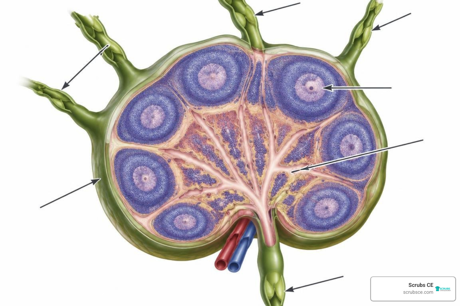
Each node features a dense fibrous capsule that extends inward as trabeculae, dividing the interior into distinct compartments:
- Cortex (Outer Zone): Houses germinal centers rich in proliferating B lymphocytes, surrounded by T cells and dendritic cells.
- Medulla (Inner Core): Contains medullary cords and sinuses populated by macrophages, plasma cells, and antibody-secreting B cells.
Fluid enters a lymph node through multiple afferent lymphatic vessels, forcing the fluid slowly through winding subcapsular and medullary sinuses. Here, resident macrophages and dendritic cells engulf foreign matter, bacteria, and cellular debris. The purified lymph then exits through only one or two efferent lymphatic vessels at the indented hilum.
Because there are more entry doors (afferent) than exit doors (efferent), fluid slows down inside the node, giving immune cells plenty of time to inspect every droplet. Lymph nodes cluster heavily in high-exposure regions:
- Cervical Nodes: Neck region; monitor lymph from the head and throat.
- Axillary Nodes: Armpits; filter drainage from the upper limbs and mammary glands.
- Inguinal Nodes: Groin; filter lymph arriving from the lower extremities and external genitalia.
How the Immune System Operates Inside the Lymphatic Network
The lymphatic system functions as the primary physical framework for immunity, providing the communication lines and deployment zones where white blood cells intercept pathogens, as outlined in the SEER Training review of lymphatic pathways and structures.
Key Immune Cells: Lymphocytes, Phagocytes, and Granulocytes
The body maintains an army of roughly $10^{12}$ lymphocytes, supported by diverse white blood cell (leukocyte) populations that circulate between blood, lymph, and tissues:
- Neutrophils (40% to 70% of total WBCs): The rapid-response phagocytes of the innate immune system. They arrive first at sites of acute infection, engulfing bacteria and releasing antimicrobial chemicals before dying to form pus.
- Lymphocytes (20% to 45% of total WBCs): The specialized orchestrators of targeted immunity:
- B Lymphocytes (B Cells): Differentiate into plasma cells that synthesize targeted antibodies. Immunoglobulin G (IgG) antibodies represent approximately 80% of all circulating antibodies in the body.
- T Lymphocytes (T Cells): Direct cellular attacks via cytotoxic T cells ($CD8^+$) or coordinate broader immune responses through helper T cells ($CD4^+$).
- Natural Killer (NK) Cells: Innate lymphocytes that identify and destroy virus-infected cells and abnormal tumor cells without prior sensitization.
- Monocytes (4% to 8% of total WBCs): Large circulating leukocytes that migrate into tissues to differentiate into voracious macrophages and dendritic cells.
- Eosinophils (1% to 4% of total WBCs): Granulocytes specialized in attacking parasitic worms and modulating allergic inflammatory reactions.
- Basophils (~1% of total WBCs): Granulocytes that release histamine and heparin to promote inflammation and recruit other immune cells to injury sites.
- Dendritic Cells: Antigen-presenting cells residing in tissues that capture foreign particles and travel through lymphatics to present them to T cells in regional nodes.
- The Complement System: A biochemical cascade composed of about 26 plasma proteins that work alongside antibodies to punch holes in bacterial membranes and flag invaders for destruction.
Innate vs. Adaptive Immunity in Lymphatic Tissues
Our immune response divides into two coordinated branches that cooperate inside lymphoid tissues, a foundational topic explored in anatomy and physiology for healthcare workers:
| Feature | Innate Immunity (Non-Specific) | Adaptive Immunity (Specific) |
|---|---|---|
| Response Speed | Immediate (minutes to hours) | Delayed (days to weeks upon first exposure) |
| Specificity | Broad; recognizes general pathogen patterns | Highly specific; targets unique antigen epitopes |
| Memory | No immunological memory | Generates long-lived memory B and T cells |
| Key Components | Skin, mucus, neutrophils, macrophages, NK cells, complement | Helper T cells, Cytotoxic T cells, B cells, Antibodies |
| Primary Location | Epithelial barriers, connective tissues, sinuses | Germinal centers of lymph nodes, spleen, MALT |
Innate defenses form our first line of defense (physical barriers like skin and mucous membranes) and second line of defense (phagocytes, inflammation, and fever). When innate cells cannot clear an infection on their own, dendritic cells pick up antigen fragments, migrate into local lymph nodes, and activate adaptive T and B lymphocytes for a targeted response.
Common Lymphatic Pathologies and How to Keep Your System Healthy
When the lymphatic system encounters structural damage, genetic abnormalities, or overwhelming infections, fluid dynamics and immune defense can falter, as cataloged in the MedlinePlus medical encyclopedia resource on lymphatic structures.
Clinical Disorders: From Lymphedema to Lymphadenopathy
- Lymphedema: Chronic tissue swelling caused by obstructed or damaged lymphatic drainage. Primary lymphedema stems from congenital vessel malformations, whereas secondary lymphedema arises from external damage, such as surgical lymph node dissection during cancer staging, radiation therapy scarring, or traumatic injury.
- Lymphadenopathy: Abnormal enlargement of lymph nodes, typically reflecting an active immune response to local infection (reactive hyperplasia), systemic autoimmune disease, or malignant infiltration.
- Lymphangitis: An acute bacterial infection (frequently Streptococcus or Staphylococcus) causing inflammation of lymphatic channels, visible as tender, erythematous red streaks tracking up an extremity toward regional nodes.
- Lymphoma: Primary malignancies of lymphoid tissue, categorized into:
- Hodgkin Lymphoma: Characterized histologically by the presence of giant, multinucleated Reed-Sternberg cells.
- Non-Hodgkin Lymphoma: A diverse group of B-cell or T-cell malignancies lacking Reed-Sternberg cells.
- Lymphatic Filariasis: A tropical parasitic infection caused by mosquito-borne microscopic roundworms (Wuchereria bancrofti) that lodge in lymphatic vessels, causing severe chronic obstruction and profound tissue hypertrophy known as elephantiasis. Exploring how these conditions alter cellular architecture is a core element of pathological anatomy principles.
Daily Habits to Support Lymphatic Drainage
Because the lymphatic system relies on external mechanical forces to move fluid, proactive daily habits can help support natural drainage:
- Regular Physical Exercise: Dynamic aerobic activities such as walking, swimming, and rebounding create rhythmic skeletal muscle contractions that pump lymph through peripheral vessels.
- Diaphragmatic Deep Breathing: Deep belly breathing causes significant pressure shifts between the abdominal and thoracic cavities, drawing fluid upward through the thoracic duct.
- Adequate Hydration: Drinking sufficient water maintains healthy blood viscosity and keeps interstitial fluids moving smoothly through tissue capillaries.
- Dry Brushing and Manual Lymphatic Drainage: Gentle, superficial skin brushing directed toward the heart can stimulate initial lymphatic capillaries in people prone to mild fluid retention.
- Compression Garments: Medical-grade graduated compression sleeves or stockings provide external pressure to assist venous and lymphatic return in individuals managing chronic venous insufficiency or secondary lymphedema.
Frequently Asked Questions About the Lymphatic System
What is the difference between lymph and blood plasma?
Blood plasma is the fluid component of whole blood, rich in dissolved proteins (such as albumin, fibrinogen, and globulins), electrolytes, nutrients, and suspended red blood cells, white blood cells, and platelets.
Interstitial fluid is plasma that has filtered out of capillaries into tissue spaces, leaving behind large proteins and blood cells. Once this interstitial fluid enters lymphatic capillaries, it is called lymph. Lymph lacks red blood cells and platelets, has a lower protein concentration than plasma, contains elevated numbers of lymphocytes, and carries absorbed lipids (chyle) when collected from the digestive tract.
Why do lymph nodes swell when you are sick?
When bacteria, viruses, or foreign antigens enter your tissues, local dendritic cells and fluid carry them straight to regional lymph nodes. Resident lymphocytes with matching receptors recognize these antigens and begin dividing rapidly in germinal centers (a process called clonal expansion).
This sudden increase in white blood cells, combined with the arrival of additional immune cells and localized fluid congestion, causes the node to enlarge and feel tender to the touch.
Does the lymphatic system have its own central pump?
No. Unlike the cardiovascular system, which relies on the heart to push blood under high pressure, the lymphatic system has no central pump. It is a low-pressure drainage network that depends entirely on external mechanical forces:
- Contractions of nearby skeletal muscles
- Pressure changes created by the diaphragm during respiration
- Rhythmic pulsations of adjacent arteries
- Contractions of smooth muscle within the walls of larger collecting ducts
- Series of internal one-way valves that prevent backflow
Conclusion
The lymphatic system serves two equally vital roles: it maintains fluid balance by returning 3 liters of filtered plasma to the bloodstream every day, and it acts as the primary command network for immune defense. From microscopic endothelial minivalves to specialized filtration hubs in lymph nodes and the spleen, this system keeps our internal environment clean, balanced, and protected against disease.
For medical imaging technologists, nursing professionals, and healthcare students, mastering these anatomical pathways is essential for evaluating clinical scans, managing patient edema, and identifying signs of pathology. At Scrubs CE, we provide high-quality, self-paced continuing education courses with instant certificates to help healthcare professionals meet licensure requirements and advance their careers.
If you are a radiologic technologist seeking credits accepted by the ARRT® (the AMERICAN REGISTRY OF RADIOLOGIC TECHNOLOGISTS®) or looking to fulfill state requirements, explore our comprehensive Anatomy & Physiology for Dummies 3rd Ed course today to refresh your foundational knowledge and keep your clinical expertise current.