

Core Foundations: Connecting Structure, Function, and Homeostasis
Busy students can learn anatomy and physiology conveniently by using active recall, spaced repetition, and bite-sized visual tools while anchoring new terms in the body’s biological hierarchy. Instead of memorizing endless anatomical lists, break the subject into logical levels: chemical foundations, cells, tissues, organs, organ systems, and the whole organism. This structure gives every bone, vessel, and organ a logical home and makes study sessions faster and more flexible.
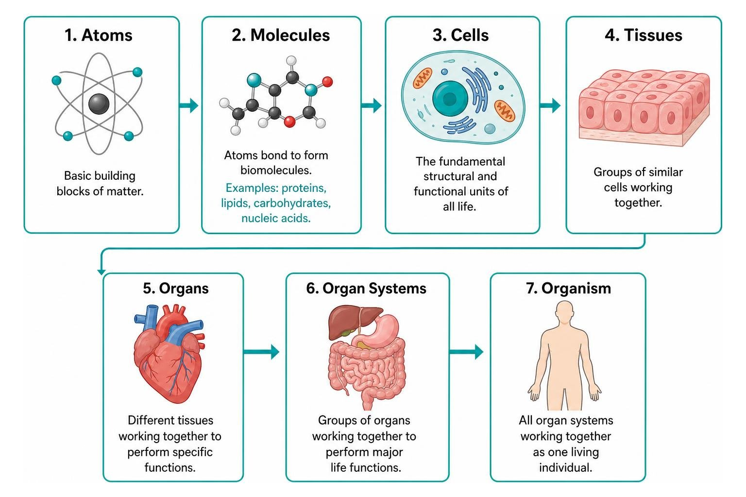
At the chemical level, basic atoms like carbon, hydrogen, and oxygen bond to form complex biomolecules, including proteins, lipids, carbohydrates, and nucleic acids. These molecules assemble into cells—the fundamental structural and functional units of all life. Within each cell, specialized organelles carry out distinct survival tasks:
- Nucleus: Houses genetic DNA, serving as the master control center for protein synthesis and cell division.
- Mitochondria: The power plants that generate adenosine triphosphate (ATP) through cellular respiration to fuel biological work.
- Endoplasmic Reticulum (ER): Synthesizes proteins (rough ER, studded with ribosomes) and lipids while neutralizing toxins (smooth ER).
- Golgi Apparatus: Modifies, sorts, and packages macromolecules for secretion or internal delivery.
- Lysosomes: Contain digestive enzymes that break down cellular debris, waste, and foreign invaders.
- Plasma Membrane: A selective phospholipid bilayer regulating nutrient entry and waste removal via diffusion, active transport, and osmosis.
When specialized cells unite to perform shared tasks, they create tissues, which then form functional organs, integrated organ systems, and ultimately the complete human organism.
| Biological Level | Structural Characteristics | Primary Physiological Function | Everyday Analogy |
|---|---|---|---|
| Chemical Level | Atoms, ions, and biomolecules (proteins, ATP, DNA) | Provides molecular machinery for metabolic reactions | Individual construction screws and electrical wires |
| Cellular Level | Organelles enclosed in a semi-permeable membrane | Carries out core life processes like energy generation | Specialized workers on an assembly line |
| Tissue Level | Sheets or clusters of similar, specialized cells | Provides unified action (contraction, covering, support) | Specialized trade departments in a factory |
| Organ Level | Distinct anatomical units composed of 2+ tissue types | Executes complex bodily operations (e.g., pumping blood) | A complete machine running a specific operation |
| Organ System Level | Interconnected organs working in tandem | Sustains major life functions (e.g., gas exchange) | An entire production facility maintaining output |
| Organism Level | All eleven systems functioning in coordinated balance | Maintains independent biological life and reproduction | A fully operating corporate enterprise |
The Interdependence of Anatomy and Physiology
Anatomy (the structural map) and physiology (the mechanical and chemical manual) are two sides of the exact same coin. Form always reflects function. For instance, the wafer-thin simple squamous epithelium lining our pulmonary alveoli exists specifically to allow oxygen and carbon dioxide to diffuse rapidly into surrounding capillaries. If the lungs were constructed from thick, multi-layered stratified skin cells, we would suffocate within minutes.
Understanding these foundational body manual concepts transforms rote memorization into intuitive comprehension. When you study the shape of a bone joint, the muscular orientation of the stomach wall, or the layered electrical conduction system of the heart, always ask yourself: Why is it shaped this way, and what job does that shape make possible?
From Cells and Tissues to Organ Systems
Every single organ in our body is constructed from four primary tissue types, each tailored for a distinct biological role:
- Epithelial Tissue: Covers external body surfaces, lines internal cavities, and forms glands. It serves as an active barrier for protection, absorption, filtration, and secretion.
- Connective Tissue: The most abundant tissue variety, connecting and supporting structures throughout the body. It includes loose areolar tissue, dense fibrous ligaments, adipose cushions, rigid bone matrix, flexible cartilage, and liquid blood.
- Muscle Tissue: Specialized contractile tissue responsible for generating mechanical movement and heat. It divides into skeletal muscle (voluntary, striated), cardiac muscle (involuntary, branching, striated), and smooth muscle (involuntary, non-striated, lining visceral hollow organs).
- Nervous Tissue: Composed of excitability-driven neurons and supportive glial cells. It coordinates internal electrical communication and processes external sensory stimuli.
These tissues combine to construct the eleven functional body systems. For students who want simplified explanations of these structures, learning these simplified healthcare fundamentals builds a solid groundwork for clinical practice.
Maintaining Homeostatic Equilibrium
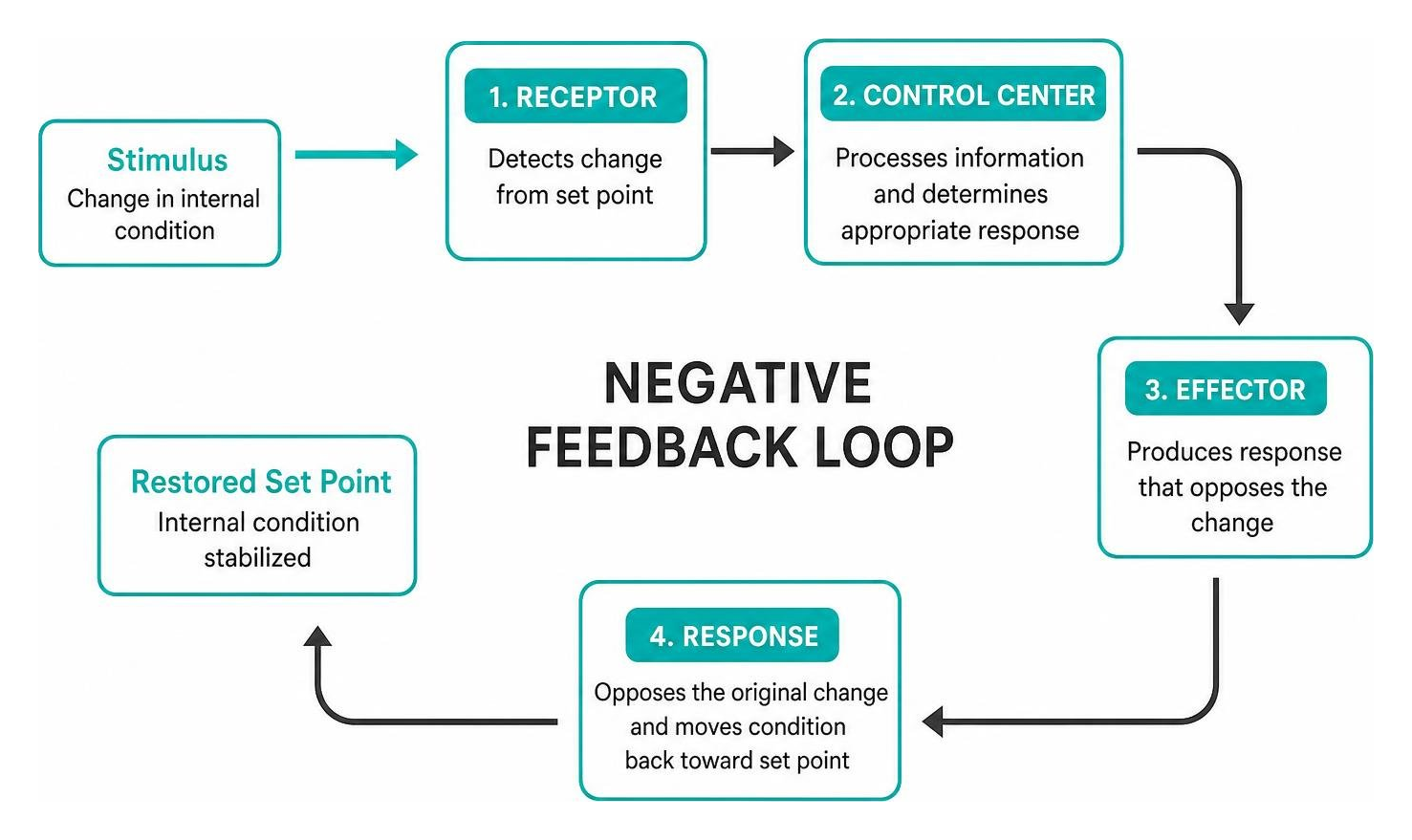
Homeostasis is the dynamic, self-regulating process by which the body preserves internal physical and chemical stability despite constant external fluctuations. Core variables—such as a body temperature near 98.6°F (37°C), arterial blood pH between 7.35 and 7.45, and resting blood glucose levels—must remain inside narrow physiological limits to support cellular survival.
The body monitors and adjusts these parameters through automated feedback mechanisms:
- Negative Feedback Loops: The primary homeostatic mechanism, where the biological response opposes or reverses the original stimulus. When your core temperature spikes on a hot afternoon, thermoreceptors alert the hypothalamus, triggering peripheral vasodilation and sweat production to shed heat. Once cooling occurs, sweating stops.
- Positive Feedback Loops: Less common pathways where the physiological response enhances or accelerates the initial deviation until a definitive terminal event occurs. Classic examples include uterine oxytocin release intensifying contractions during labor and platelet aggregation cascade sealing an injured blood vessel during clotting.
Mastering the Map: Directional Terms, Planes, and Body Regions
Navigating the human body requires a universal language. Just as a navigator uses cardinal compass coordinates, medical and imaging professionals rely on standard anatomical terms to prevent costly communication errors.
Standard Anatomical Position and Planes
All anatomical descriptions assume a universally accepted baseline known as the anatomical position. In this standard stance, the subject stands completely upright, facing forward toward the observer, with feet flat and parallel on the floor, upper limbs resting at the sides, and palms facing outward with thumbs pointing away from the body.
To view internal structural relationships, we mentally slice the body using three fundamental anatomical planes:
- Sagittal Plane: Runs vertically, dividing the body or an organ into right and left portions. A midsagittal (median) plane divides the body into equal symmetrical halves, whereas a parasagittal plane creates unequal sections.
- Coronal (Frontal) Plane: Runs vertically from ear to ear, slicing the body into anterior (front) and posterior (back) portions.
- Transverse (Horizontal/Axial) Plane: Cuts horizontally across the body axis, dividing the subject into superior (upper) and inferior (lower) portions. This is the primary cross-sectional plane generated by computed tomography (CT) scanners in diagnostic Radiology.
Directional pairs allow precise location mapping:
- Anterior (Ventral) vs. Posterior (Dorsal): Front vs. back of the body.
- Superior (Cranial) vs. Inferior (Caudal): Closer to the head vs. closer to the feet.
- Medial vs. Lateral: Closer to the midline vs. farther toward the outer edge.
- Proximal vs. Distal: Closer to the point of limb attachment vs. farther down the extremity.
- Superficial vs. Deep: Near the body surface vs. deeper inside internal tissue layers.
Regional Terms and Body Cavities
Internal organs (viscera) are compartmentalized within closed body cavities that shield structures from shock and allow movement without friction:
- Dorsal (Posterior) Body Cavity: Subdivided into the cranial cavity (enclosing the brain within the skull) and the vertebral (spinal) cavity (encasing the spinal cord).
- Ventral (Anterior) Body Cavity: Divided by the muscular diaphragm into:
- Thoracic Cavity: Houses the lateral pleural cavities (lungs) and the central mediastinum, which encloses the pericardial cavity containing the heart, esophagus, and trachea.
- Abdominopelvic Cavity: Extends from the diaphragm to the pelvic basin. For clinical exams, healthcare providers divide the abdomen into four quadrants (Right Upper Quadrant, Left Upper Quadrant, Right Lower Quadrant, Left Lower Quadrant) or nine anatomical regions to pinpoint pathology, such as locating appendix inflammation in the right lower quadrant.
Convenient Anatomy and Physiology Learning: A Step-by-Step Blueprint
Studying anatomy does not require locking yourself in a library with an eleven-hundred-page text. High-yield, self-paced learning centers on structuring your study time into focused, bite-sized intervals. Many students use structured study guides to quickly break down tough topics into plain English.
Active Recall and Spaced Repetition Tactics
Rereading highlighted textbook paragraphs creates a false sense of mastery. Real retention happens when your brain works to pull facts out of memory:
- Spaced Retrieval Schedules: Rather than cramming for six straight hours before an exam, study a single organ system for 30 minutes a day over five days. Revisit challenging concepts at expanding intervals (Day 1, Day 3, Day 7, Day 14).
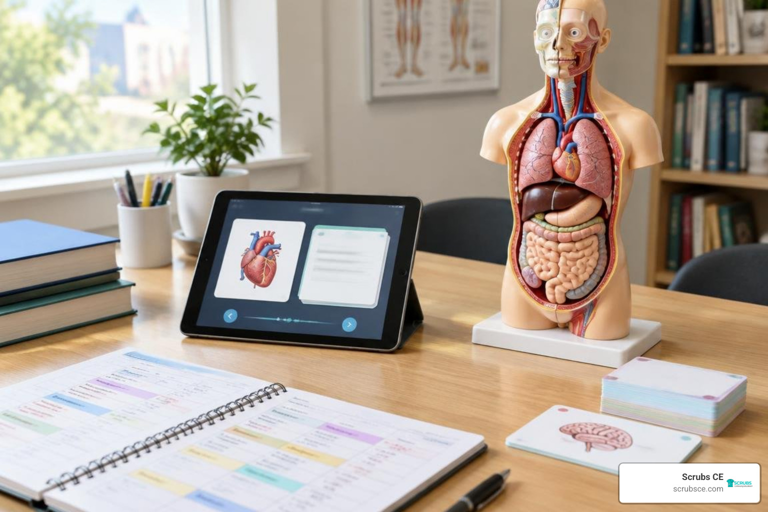
- Anatomy Flashcards with Occlusion: Use digital flashcards to conceal anatomical labels on unlabeled line drawings. Force yourself to name the muscle insertion, nerve innervation, or vascular branch out loud before flipping the card.
- Medical Etymology Deconstruction: Anatomy terminology is built largely on Greek and Latin roots. Deconstruct complex words into manageable pieces:
- Cardio- (heart) + -myo- (muscle) + -pathy- (disease) = Disease of the heart muscle.
- Chondro- (cartilage) + -cyte (cell) = Mature cartilage cell.
- Hepato- (liver) + -itis (inflammation) = Liver inflammation.
- Blank-Page Concept Mapping: Close your books and draw the systemic circulatory circuit or the parts of a kidney nephron entirely from memory. Once finished, open your notes in a different colored pen to immediately spot and correct knowledge gaps.
Leveraging Modern Visual and Digital Tools
Pairing written notes with visual aids creates two separate memory pathways in the brain:
- Interactive 3D Virtual Atlases: Tools like 3D interactive models allow you to digitally peel away fascia, muscles, and bones layer by layer to see exact spatial relationships.
- Hybrid Text and Question Banks: Comprehensive resources with online chapter quizzes give you instant scoring feedback so you know which systems need a quick review.
- Standard Atlases and Quick Guides: Comprehensive college references provide layered illustrations, clinical sidebars, and microbiome overviews that connect microstructures to real clinical scenarios. For a broader overview of the field, see anatomy.
Clinical Context: Pathophysiology and Real-World Application
Anatomy comes alive the moment you see what happens when healthy structures break down. Exploring disease mechanisms (pathophysiology) shows how subtle changes in tissue anatomy produce classic medical symptoms.

Convenient Anatomy and Physiology Learning for Exam Prep
Whether you are preparing for college midterms, national registry boards, or nursing and allied health certifications, efficient A&P prep relies on targeted review.
Understanding your professional licensure requirements helps you focus on high-yield, clinically relevant material. If you are shopping for study materials, make sure you are selecting the best edition with up-to-date terminology, online test banks, and clear visual diagrams.
When taking timed multiple-choice exams, watch for classic board exam patterns:
- Identify the structural “odd-one-out” in tissue classifications.
- Trace fluids or electrical pathways step-by-step (e.g., following blood flow through heart valves, or electrical conduction from the SA node to Purkinje fibers).
- Predict how hormone changes alter lab values (such as parathyroid hormone driving up blood calcium levels).
Convenient Anatomy and Physiology Learning in Clinical Practice
In healthcare, structural anatomy is a daily working tool. Radiologic technologists rely on anatomical landmarks to position patients accurately for diagnostic imaging, while nurses assess physical signs to catch early organ dysfunction.
Here is how common clinical pathologies highlight underlying anatomical and physiological principles:
- Cardiovascular System (Myocardial Infarction): An atherosclerotic plaque ruptures inside a coronary artery, blocking blood flow to downstream cardiac muscle tissue. Deprived of oxygen, the tissue cannot produce ATP, cardiac cells die, and normal myocardial contraction fails.
- Respiratory System (COPD and Emphysema): Chronic irritation destroys alveolar wall elasticity, reducing the surface area available for gas exchange. Air becomes trapped in the lungs, leading to barrel-chest anatomy, dyspnea, and systemic respiratory acidosis.
- Nervous System (Multiple Sclerosis): Autoimmune destruction targets the protective myelin sheath surrounding central nervous system axons. Without proper insulation, electrical action potential conduction slows, producing muscle weakness, vision loss, and coordination deficits.
- Endocrine System (Type 1 Diabetes Mellitus): Autoimmune destruction wipes out insulin-producing beta cells in pancreatic islets of Langerhans. Without insulin to facilitate cellular glucose uptake, glucose builds up in the bloodstream while body cells starve for usable metabolic fuel.
Gaining real clinical confidence means connecting standard structure to pathology. You can explore these mechanisms in our guide to pathological anatomy insights and review everyday medical imaging concepts in our primer on imaging anatomy fundamentals.
Frequently Asked Questions About Studying Anatomy and Physiology
How can beginners learn anatomy and physiology quickly without getting overwhelmed?
Focus on mastering the big picture before memorizing every tiny detail. Start by understanding what an entire organ system does, then zoom in to learn its main organs, tissue layers, and cellular processes.
Study in focused 25-to-30-minute intervals using active recall and dual-coding techniques (pairing written descriptions with labeled diagrams) instead of rereading full chapters for hours.
What is the most effective way to memorize anatomical terminology?
The fastest way to master anatomical vocabulary is to learn common Greek and Latin prefixes, roots, and suffixes. Because most medical terms are combinations of descriptive word parts (such as sub- meaning below, costal referring to ribs, and -algia meaning pain), learning 50 common roots unlocks the meaning of hundreds of complex terms without brute-force memorization.
Combine this root-word approach with digital flashcards and directional body maps.
Why is understanding pathophysiology critical when learning normal anatomy?
Pathophysiology acts as a real-world stress test for normal anatomy and physiology. When you see how a diseased or damaged structure impairs a patient’s health—like how a narrowed heart valve causes fluid backup into the lungs—the normal structural layout and its physiological purpose become far easier to remember.
Clinical context turns abstract science into practical, memorable knowledge.
Conclusion
Mastering human anatomy and physiology does not require endless, stressful cramming. By anchoring your study habits around biological hierarchy, homeostatic feedback loops, and directional language, you can break down complex medical topics into clear, intuitive concepts. Combining active recall techniques, 3D visual models, and clinical case examples makes study time manageable and helps you retain what you learn throughout your entire healthcare career.
At Scrubs CE, we support busy healthcare students, radiologic technologists, and medical professionals with convenient, affordable online continuing education. Whether you are earning credits for your state license or meeting American Registry of Radiologic Technologists® (ARRT®) requirements, our self-paced courses and instant certificates make professional development simple, flexible, and accessible.
Take control of your learning schedule today. Discover how simple, flexible continuing education can be by exploring our comprehensive Anatomy & Physiology for Dummies Course at Scrubs CE!